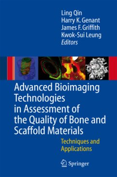
Advanced Bioimaging Technologies in Assessment of the Quality of Bone and Scaffold Materials
Techniques and Applications
Herausgegeben:Qin, L.; Genant, Harry K.; Griffith, J.F.; Leung, K.S.
Advanced Bioimaging Technologies in Assessment of the Quality of Bone and Scaffold Materials
Techniques and Applications
Herausgegeben:Qin, L.; Genant, Harry K.; Griffith, J.F.; Leung, K.S.
- Broschiertes Buch
- Merkliste
- Auf die Merkliste
- Bewerten Bewerten
- Teilen
- Produkt teilen
- Produkterinnerung
- Produkterinnerung
This book provides a perspective on the current status of bioimaging technologies developed to assess the quality of musculoskeletal tissue with an emphasis on bone and cartilage. It offers evaluations of scaffold biomaterials developed for enhancing the repair of musculoskeletal tissues. These bioimaging techniques include micro-CT, nano-CT, pQCT/QCT, MRI, and ultrasound, which provide not only 2-D and 3-D images of the related organs or tissues, but also quantifications of the relevant parameters. The advance bioimaging technologies developed for the above applications are also extended by…mehr
Andere Kunden interessierten sich auch für
![Imaging of the Hip & Bony Pelvis Imaging of the Hip & Bony Pelvis]() Imaging of the Hip & Bony Pelvis164,99 €
Imaging of the Hip & Bony Pelvis164,99 €![Tissue Characterization in MR Imaging Tissue Characterization in MR Imaging]() Tissue Characterization in MR Imaging77,99 €
Tissue Characterization in MR Imaging77,99 €![Tissue Engineering Tissue Engineering]() John P. Fisher (ed.)Tissue Engineering153,99 €
John P. Fisher (ed.)Tissue Engineering153,99 €![Radiology of Osteoporosis Radiology of Osteoporosis]() Radiology of Osteoporosis82,99 €
Radiology of Osteoporosis82,99 €![Analysis and Assessment of Cardiovascular Function Analysis and Assessment of Cardiovascular Function]() Analysis and Assessment of Cardiovascular Function75,99 €
Analysis and Assessment of Cardiovascular Function75,99 €![Deformable Models Deformable Models]() Deformable Models149,99 €
Deformable Models149,99 €![Methods of External Hyperthermic Heating Methods of External Hyperthermic Heating]() Methods of External Hyperthermic Heating39,99 €
Methods of External Hyperthermic Heating39,99 €-
-
-
This book provides a perspective on the current status of bioimaging technologies developed to assess the quality of musculoskeletal tissue with an emphasis on bone and cartilage. It offers evaluations of scaffold biomaterials developed for enhancing the repair of musculoskeletal tissues. These bioimaging techniques include micro-CT, nano-CT, pQCT/QCT, MRI, and ultrasound, which provide not only 2-D and 3-D images of the related organs or tissues, but also quantifications of the relevant parameters. The advance bioimaging technologies developed for the above applications are also extended by incorporating imaging contrast-enhancement materials. Thus, this book will provide a unique platform for multidisciplinary collaborations in education and joint R&D among various professions, including biomedical engineering, biomaterials, and basic and clinical medicine.
Produktdetails
- Produktdetails
- Verlag: Springer / Springer Berlin Heidelberg / Springer, Berlin
- Artikelnr. des Verlages: 978-3-642-07955-9
- Softcover reprint of hardcover 1st edition 2007
- Seitenzahl: 716
- Erscheinungstermin: 14. Oktober 2010
- Englisch
- Abmessung: 235mm x 155mm x 39mm
- Gewicht: 1089g
- ISBN-13: 9783642079559
- ISBN-10: 3642079555
- Artikelnr.: 32057987
- Herstellerkennzeichnung
- Springer-Verlag GmbH
- Tiergartenstr. 17
- 69121 Heidelberg
- ProductSafety@springernature.com
Perspectives of Advances in Musculoskeletal and Scaffold Biomaterial Imaging Technologies and Applications.- Perspectives on Advances in Bone Imaging for Osteoporosis.- Bone Structure and Biomechanical Analyses Using Imaging and Simulation Technology.- Imaging Technologies for Orthopaedic Visualization and Simulation.- In-Vivo Bone Mineral Density and Structures in Humans: From Isotom Over Densiscan to Xtreme-CT.- Calibration of Micro-CT Data for Quantifying Bone Mineral and Biomaterial Density and Microarchitecture.- Repositioning of the Region of Interest in the Radius of the Growing Child in Follow-up Measurements by pQCT.- Non-invasive Bone Quality Assessment Using Quantitative Ultrasound Imaging and Acoustic Parameters.- Cortical Bone Mineral Status Evaluated by pQCT, Quantitative Backscattered Electron Imaging and Polarized Light Microscopy.- High-Fidelity Histologic Three-Dimensional Analysis of Bone and Cartilage.- Application of Laser Scanning Confocal Microscopy in Musculoskeletal Research.- Fiber-optic Nano-biosensors and Near-Field Scanning Optical Microscopy for Biological Imaging.- Changes of Biological Function of Bone Cells and Effect of Anti-osteoporosis Agents on Bone Cells.- Bone Histomorphometry in Various Metabolic Bone Diseases Studied by Bone Biopsy in China.- Cell Traction Force Microscopy.- Contrast-Enhanced Micro-CT Imaging of Soft Tissues.- Materials Selection and Scaffold Fabrication for Tissue Engineering in Orthopaedics.- Quantification of Porosity, Connectivity and Material Density of Calcium Phosphate Ceramic Implants Using Micro-Computed Tomography.- Bone Densitometries in Assessing Bone Mineral and Structural Profiles in Patients with Adolescent Idiopathic Scoliosis.- Application of Nano-CT and High-Resolution Micro-CT to Study Bone Quality and Ultrastructure, Scaffold Biomaterials and Vascular Networks.- Bio-imaging Technologies in Studying Bone-Biomaterial Interface: Applications in Experimental Spinal Fusion Model.- Assessment of Bone, Cartilage, Tendon and Bone Cells by Confocal Laser Scanning Microscopy.- Specific Applications of Advances in Musculoskeletal and Scaffold Biomaterial Imaging Technologies.- TEM Study of Bone and Scaffold Materials.- Material and Structural Basis of Bone Fragility: A Rational Approach to Therapy.- Application of Micro-CT and MRI in Clinical and Preclinical Studies of Osteoporosis and Related Disorders.- CT-Based Microstructure Analysis for Assessment of Bone Fragility.- Discrimination of Contributing Factors to Bone Fragility Using vQCT In Vivo.- Osteoporosis Research with the vivaCT40.- Mechanical Properties of Vertebral Trabeculae with Ageing Evaluated with Micro-CT.- MRI Evaluation of Osteoporosis.- Multiple Bio-imaging Modalities in Evaluation of Epimedium-Derived Phytoestrogenic Fraction for Prevention of Postmenopausal Osteoporosis.- Areal and Volumetric Bone Densitometry in Evaluation of Tai Chi Chuan Exercise for Prevention of Postmenopausal Osteoporosis.- Enhancement of Osteoporotic Bone Using Injectable Hydroxyapatite in OVX Goats Evaluated by Multi-imaging Modalities.- Quality of Healing Compared Between Osteoporotic Fracture and Normal Traumatic Fracture.- Monitoring Fracture Healing Using Digital Radiographies.- Fracture Callus Under Anti-resorptive Agent Treatment Evaluated by pQCT.- Volumetric Measurement of Osteonecrotic Femoral Head Using Computerized MRI and Prediction For Its Mechanical Properties.- Biomedical Engineering in Surgical Repair of Osteonecrosis: the Role of Imaging Technology.- Contrast-Enhanced MRI and Micro-CT Adopted for Evaluationof a Lipid-Lowering and Anticoagulant Herbal Epimedium-Derived Phytoestrogenic Extract for Prevention of Steroid-Associated Osteonecrosis.- Nanomechanics of Bone and Bioactive Bone-Cement Interfaces.- Subchondral Bone Microarchitecture Changes in Animal Models of Arthritis.- Microarchitectural Adaptations of Primary Osteoarthrotic Subchondral Bone.- Ultrasonic Characterization of Dynamic Depth-Dependent Biomechanical Properties of Articular Cartilage.- Mechanical Property of Trabecular Bone of the Femoral Heads from Osteoarthritis and Osteoporosis Patients.
Perspectives of Advances in Musculoskeletal and Scaffold Biomaterial Imaging Technologies and Applications.- Perspectives on Advances in Bone Imaging for Osteoporosis.- Bone Structure and Biomechanical Analyses Using Imaging and Simulation Technology.- Imaging Technologies for Orthopaedic Visualization and Simulation.- In-Vivo Bone Mineral Density and Structures in Humans: From Isotom Over Densiscan to Xtreme-CT.- Calibration of Micro-CT Data for Quantifying Bone Mineral and Biomaterial Density and Microarchitecture.- Repositioning of the Region of Interest in the Radius of the Growing Child in Follow-up Measurements by pQCT.- Non-invasive Bone Quality Assessment Using Quantitative Ultrasound Imaging and Acoustic Parameters.- Cortical Bone Mineral Status Evaluated by pQCT, Quantitative Backscattered Electron Imaging and Polarized Light Microscopy.- High-Fidelity Histologic Three-Dimensional Analysis of Bone and Cartilage.- Application of Laser Scanning Confocal Microscopy in Musculoskeletal Research.- Fiber-optic Nano-biosensors and Near-Field Scanning Optical Microscopy for Biological Imaging.- Changes of Biological Function of Bone Cells and Effect of Anti-osteoporosis Agents on Bone Cells.- Bone Histomorphometry in Various Metabolic Bone Diseases Studied by Bone Biopsy in China.- Cell Traction Force Microscopy.- Contrast-Enhanced Micro-CT Imaging of Soft Tissues.- Materials Selection and Scaffold Fabrication for Tissue Engineering in Orthopaedics.- Quantification of Porosity, Connectivity and Material Density of Calcium Phosphate Ceramic Implants Using Micro-Computed Tomography.- Bone Densitometries in Assessing Bone Mineral and Structural Profiles in Patients with Adolescent Idiopathic Scoliosis.- Application of Nano-CT and High-Resolution Micro-CT to Study Bone Quality and Ultrastructure, Scaffold Biomaterials and Vascular Networks.- Bio-imaging Technologies in Studying Bone-Biomaterial Interface: Applications in Experimental Spinal Fusion Model.- Assessment of Bone, Cartilage, Tendon and Bone Cells by Confocal Laser Scanning Microscopy.- Specific Applications of Advances in Musculoskeletal and Scaffold Biomaterial Imaging Technologies.- TEM Study of Bone and Scaffold Materials.- Material and Structural Basis of Bone Fragility: A Rational Approach to Therapy.- Application of Micro-CT and MRI in Clinical and Preclinical Studies of Osteoporosis and Related Disorders.- CT-Based Microstructure Analysis for Assessment of Bone Fragility.- Discrimination of Contributing Factors to Bone Fragility Using vQCT In Vivo.- Osteoporosis Research with the vivaCT40.- Mechanical Properties of Vertebral Trabeculae with Ageing Evaluated with Micro-CT.- MRI Evaluation of Osteoporosis.- Multiple Bio-imaging Modalities in Evaluation of Epimedium-Derived Phytoestrogenic Fraction for Prevention of Postmenopausal Osteoporosis.- Areal and Volumetric Bone Densitometry in Evaluation of Tai Chi Chuan Exercise for Prevention of Postmenopausal Osteoporosis.- Enhancement of Osteoporotic Bone Using Injectable Hydroxyapatite in OVX Goats Evaluated by Multi-imaging Modalities.- Quality of Healing Compared Between Osteoporotic Fracture and Normal Traumatic Fracture.- Monitoring Fracture Healing Using Digital Radiographies.- Fracture Callus Under Anti-resorptive Agent Treatment Evaluated by pQCT.- Volumetric Measurement of Osteonecrotic Femoral Head Using Computerized MRI and Prediction For Its Mechanical Properties.- Biomedical Engineering in Surgical Repair of Osteonecrosis: the Role of Imaging Technology.- Contrast-Enhanced MRI and Micro-CT Adopted for Evaluationof a Lipid-Lowering and Anticoagulant Herbal Epimedium-Derived Phytoestrogenic Extract for Prevention of Steroid-Associated Osteonecrosis.- Nanomechanics of Bone and Bioactive Bone-Cement Interfaces.- Subchondral Bone Microarchitecture Changes in Animal Models of Arthritis.- Microarchitectural Adaptations of Primary Osteoarthrotic Subchondral Bone.- Ultrasonic Characterization of Dynamic Depth-Dependent Biomechanical Properties of Articular Cartilage.- Mechanical Property of Trabecular Bone of the Femoral Heads from Osteoarthritis and Osteoporosis Patients.

