All you need to know about breast calcifications
Versandkostenfrei!
Versandfertig in 6-10 Tagen
29,99 €
inkl. MwSt.

PAYBACK Punkte
15 °P sammeln!
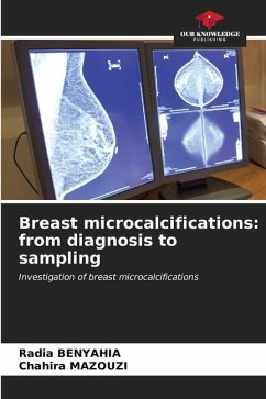
Microcalcifications are an indirect sign of the mammary pathological process. They need to be studied in detail, together with other mammographic findings, before they can be classified. Their analysis includes a description of their morphology, number and distribution, the presence or absence of associated signs, and their evolution over time. They are then classified according to the context and the patient's history. Suspected microcalcifications should always be biopsy-checked under imaging before any therapeutic decision is taken. The occurrence of postoperative calcifications may pose pr...
Microcalcifications are an indirect sign of the mammary pathological process. They need to be studied in detail, together with other mammographic findings, before they can be classified. Their analysis includes a description of their morphology, number and distribution, the presence or absence of associated signs, and their evolution over time. They are then classified according to the context and the patient's history. Suspected microcalcifications should always be biopsy-checked under imaging before any therapeutic decision is taken. The occurrence of postoperative calcifications may pose problems of differential diagnosis between cytosteatonecrosis and recurrence.This clear, didactic book, richly illustrated with demonstrative diagrams, is intended for radiologists.


