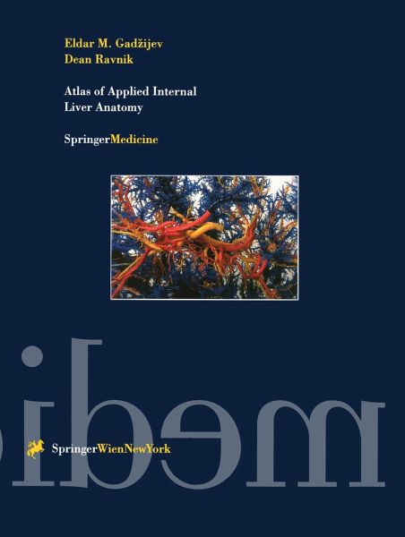
Atlas of Applied Internal Liver Anatomy
Versandkostenfrei!
Versandfertig in 1-2 Wochen
214,99 €
inkl. MwSt.
Weitere Ausgaben:

PAYBACK Punkte
107 °P sammeln!
Following a brief outline of the technology involved in preparing casts as well as the types of casts made, this atlas devotes a whole section to three blood vessel systems (portal, arterial and venous) and the biliary system. Apart from normal anatomy, it presents all other major structural variations together with the liver blood vessel structures and bile ducts of each liver segment. The last section shows correlations between systems as well as the division of the liver into sectors and segments bas ed on relations among portal pedicles and hepatic veins.The arteries, veins, and bile ducts...
Following a brief outline of the technology involved in preparing casts as well as the types of casts made, this atlas devotes a whole section to three blood vessel systems (portal, arterial and venous) and the biliary system. Apart from normal anatomy, it presents all other major structural variations together with the liver blood vessel structures and bile ducts of each liver segment. The last section shows correlations between systems as well as the division of the liver into sectors and segments bas ed on relations among portal pedicles and hepatic veins.
The arteries, veins, and bile ducts were each injected with a different colored dye to produce around 100 striking photographs of liver casts. This close collaboration between a surgeon and an anatomist ensures that the atlas is ideally suited for surgeons specialising in the liver and biliary system, as well as for general surgeons faced with liver trauma and who have to decide on appropriate treatment. Can also be used by those involved in training to present liver structures to students and junior colleagues. The aim of this Atlas is to present the three-dimensional arrangement of the liver structures, which should be familiar to those who diagnose and treat diseases of the liver, particularly in an era when the methods of diagnostic imaging and surgical treatment are becoming increasingly sophisticated. For this purpose a series of corrosive preparations of the blood vessels and bile ducts of the liver was made and photographed. In addition to the normal situations, many frequent and rare variations are shown. The Atlas also shows some blood vessels that have not been adequately described or are not well-known in the reference literature, but are nevertheless of great importance in performing segmental liver resections.This Atlas takes a fresh approach to the subject. The method used allows the size, three-dimensional arrangement and structure of the blood vessels and bile ducts of the liver to be preserved. The majority of photographs were taken from the direction from which surgeons see the liver during an operation. This, together with the schematic presentations complementing most of the photographs, gives a further instructional value to the work. With colour photographs and explanatory text, the Atlas forms a basic guide to orientation inside the liver parenchyma, to understanding and diagnosing certain pathological processes and to planning surgcial procedures.
The arteries, veins, and bile ducts were each injected with a different colored dye to produce around 100 striking photographs of liver casts. This close collaboration between a surgeon and an anatomist ensures that the atlas is ideally suited for surgeons specialising in the liver and biliary system, as well as for general surgeons faced with liver trauma and who have to decide on appropriate treatment. Can also be used by those involved in training to present liver structures to students and junior colleagues. The aim of this Atlas is to present the three-dimensional arrangement of the liver structures, which should be familiar to those who diagnose and treat diseases of the liver, particularly in an era when the methods of diagnostic imaging and surgical treatment are becoming increasingly sophisticated. For this purpose a series of corrosive preparations of the blood vessels and bile ducts of the liver was made and photographed. In addition to the normal situations, many frequent and rare variations are shown. The Atlas also shows some blood vessels that have not been adequately described or are not well-known in the reference literature, but are nevertheless of great importance in performing segmental liver resections.This Atlas takes a fresh approach to the subject. The method used allows the size, three-dimensional arrangement and structure of the blood vessels and bile ducts of the liver to be preserved. The majority of photographs were taken from the direction from which surgeons see the liver during an operation. This, together with the schematic presentations complementing most of the photographs, gives a further instructional value to the work. With colour photographs and explanatory text, the Atlas forms a basic guide to orientation inside the liver parenchyma, to understanding and diagnosing certain pathological processes and to planning surgcial procedures.


