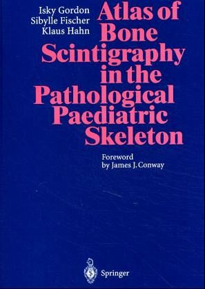
Atlas of Bone Scintigraphy in the Pathological Paediatric Skeleton
Under the Auspices of the Paediatric Committee of the European Association of Nuclear Medicine. Forew. by James J, Conway
Versandkostenfrei!
Nicht lieferbar
Weitere Ausgaben:
This atlas covers both the common and the less common pathologies affecting the paediatric skeleton, providing illustrations, teaching points, technical comments, and text discussion. Variations in the appearances of osteomyelitis are also extensively illustrated. The procedures employed to create the images presented in this volume include whole body scanning, gamma camera high resolution spot images, pin hole and SPECT. Three phase bone scans are also illustrated. Indications for the use of each procedure are dicussed. The many illustrations in this atlas offer the paediatrician, orthopaedic surgeon, radiologist and nuclear medicine physician the opportunity to compare them with their own images or with the "normal" images presented in the previous companion volume.
In diesem Atlas wird die allgemeine und spezielle pathologische Entwicklung des kindlichen Skeletts in szintigraphischen Abbildungen dargestellt. Dieses Buch unterstützt den Pädiater, Orthopäden, Radiologen und Nuklearmediziner in der täglichen Praxis. Im Vergleich der eigenen Aufnahmen mit den Abbildungen kann eine sichere Diagnose gestellt werden. Jeder Fall wird durch einen Kommentar ergänzt.


