J. Bradley Randleman
Atlas of Corneal Imaging
J. Bradley Randleman
Atlas of Corneal Imaging
- Gebundenes Buch
- Merkliste
- Auf die Merkliste
- Bewerten Bewerten
- Teilen
- Produkt teilen
- Produkterinnerung
- Produkterinnerung
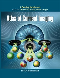
A comprehensive reference for physicians, surgeons, and trainees, Atlas of Corneal Imaging covers all aspects of corneal imaging from basic map interpretation to advanced diagnostic uses and features over 1200 illustrative images and figures representing a wide variety of devices and techniques.
Andere Kunden interessierten sich auch für
![Corneal Cross-Linking Corneal Cross-Linking]() Farhad HafeziCorneal Cross-Linking259,99 €
Farhad HafeziCorneal Cross-Linking259,99 €![Mastering Corneal Surgery Mastering Corneal Surgery]() Amar AgarwalMastering Corneal Surgery250,99 €
Amar AgarwalMastering Corneal Surgery250,99 €![Corneal Collagen Cross Linking Corneal Collagen Cross Linking]() Corneal Collagen Cross Linking81,99 €
Corneal Collagen Cross Linking81,99 €![Advances in Corneal Research Advances in Corneal Research]() Advances in Corneal Research38,99 €
Advances in Corneal Research38,99 €![Lamellar Corneal Surgery Lamellar Corneal Surgery]() Thomas JohnLamellar Corneal Surgery320,99 €
Thomas JohnLamellar Corneal Surgery320,99 €![Complications in Corneal Laser Surgery Complications in Corneal Laser Surgery]() Complications in Corneal Laser Surgery66,99 €
Complications in Corneal Laser Surgery66,99 €![Corneal Surgery Corneal Surgery]() Bruno ZuberbuhlerCorneal Surgery81,99 €
Bruno ZuberbuhlerCorneal Surgery81,99 €-
-
-
A comprehensive reference for physicians, surgeons, and trainees, Atlas of Corneal Imaging covers all aspects of corneal imaging from basic map interpretation to advanced diagnostic uses and features over 1200 illustrative images and figures representing a wide variety of devices and techniques.
Hinweis: Dieser Artikel kann nur an eine deutsche Lieferadresse ausgeliefert werden.
Hinweis: Dieser Artikel kann nur an eine deutsche Lieferadresse ausgeliefert werden.
Produktdetails
- Produktdetails
- Verlag: CRC Press
- Seitenzahl: 696
- Erscheinungstermin: 15. Juli 2022
- Englisch
- Abmessung: 286mm x 221mm x 42mm
- Gewicht: 1981g
- ISBN-13: 9781630916473
- ISBN-10: 1630916471
- Artikelnr.: 68676050
- Herstellerkennzeichnung
- Libri GmbH
- Europaallee 1
- 36244 Bad Hersfeld
- gpsr@libri.de
- Verlag: CRC Press
- Seitenzahl: 696
- Erscheinungstermin: 15. Juli 2022
- Englisch
- Abmessung: 286mm x 221mm x 42mm
- Gewicht: 1981g
- ISBN-13: 9781630916473
- ISBN-10: 1630916471
- Artikelnr.: 68676050
- Herstellerkennzeichnung
- Libri GmbH
- Europaallee 1
- 36244 Bad Hersfeld
- gpsr@libri.de
J. Bradley Randleman, MD is a Professor in the Department of Ophthalmology at the Cleveland Clinic Lerner College of Medicine of Case Western Reserve University and Staff Ophthalmologist and Co-Director of the Refractive Surgery Section at the Cole Eye Institute of the Cleveland Clinic Foundation in Cleveland, Ohio. Prior to his arrival in Cleveland, Dr. Randleman was Professor of Ophthalmology at the Keck School of Medicine of University of Southern California and Director of the Cornea & Refractive Surgery Service at the University of Southern California Roski Eye Institute in Los Angeles, California, and the Hughes Professor of Ophthalmology at Emory University and Director of the Cornea Section at the Emory Eye Center. A widely respected cornea specialist, his areas of expertise include corneal and intraocular (IOL) refractive surgical procedures, including LASIK and premium cataract and IOL surgery, complicated cataract surgery, and the management of corneal ectatic disorders. His primary research focuses on identification and management of corneal ectatic diseases, including keratoconus and postoperative ectasia after LASIK, and the avoidance, diagnosis, and management of refractive surgical complications. He has been awarded multiple research grants throughout his career, including R01 funding from the National Institutes of Health to evaluate corneal biomechanical analysis using Brillouin microscopy. Dr. Randleman received his BA degree from Columbia College at Columbia University in New York City; his MD degree from Texas Tech University School of Medicine in Lubbock, Texas, where he was elected to the Alpha Omega Alpha medical honor society in his junior year; and followed by his Ophthalmology residency and fellowship in Cornea/External Disease, and Refractive Surgery at Emory University in Atlanta, Georgia. Dr. Randleman has been awarded the Claus Dohlman Fellow Award, the inaugural Binkhorst Young Ophthalmologist Award from the American Society of Cataract and Refractive Surgery, the Kritzinger Memorial Award, Founder's Award, President's Award, and the Inaugural Recognition Award from the International Society of Refractive Surgery, and the Secretariat Award, Achievement Award, and Senior Achievement Award from the American Academy of Ophthalmology. He was named to The Power List by The Ophthalmologist in both 2018 and 2020. Dr. Randleman has served as Editor-in-Chief for the Journal of Refractive Surgery since 2011. He has authored more than 165 peer-reviewed publications in leading ophthalmology journals in addition to 40 book chapters on refractive surgery evaluation, corneal cross-linking, and management of complications with IOLs, and has authored 4 previous textbooks, Corneal Collagen Cross-Linking; Corneal Cross-Linking, Second Edition; Refractive Surgery: An Interactive Case-Based Approach; and Intraocular Lens Surgery: Selection, Complications, and Complex Cases.
Dedication Acknowledgments About the AuthorAbout the Associate Editors
Contributing Authors Foreword by Stephen D. Klyce, PhDIntroduction Chapter
1 Fundamental Concepts in Corneal ImagingMehdi Roozbahani, MD; Marcony R.
Santhiago, MD, PhD; William J. Dupps, MD, PhD;and J. Bradley Randleman, MD
Basic Definitions and Terminology Confusing Clinical Concepts Imaging
Devices Placido-Based Reflection Devices LED-Based Reflective Devices
Tomography-Based Imaging Devices Slit Scanning-Based Tomography
Scheimpflug-Based Tomographers Optical Coherence Tomography Very
High-Frequency Digital Ultrasound Aberrometers for Wavefront Analysis
Summary Chapter 2 Corneal Imaging Devices: Applications and Set Up Mehdi
Roozbahani, MD; Marcony R. Santhiago, MD, PhD; William J. Dupps, MD,
PhD;and J. Bradley Randleman, MD Basic Device Set Up Specific Imaging
Devices Placido Topography Scanning Slit Imaging (Orbscan II) Scheimpflug
Imaging (Pentacam) Dual Scheimpflug/Placido Imaging (Galilei)
Scheimpflug/Placido Imaging (Sirius) Anterior Segment Optical Coherence
Tomography Very High-Frequency Digital Ultrasound Imaging Artifacts Summary
Chapter 3 Basic Topographic Patterns and Tomographic Correlates J. Bradley
Randleman, MD; Marcony R. Santhiago, MD, PhD; and William J. Dupps, MD, PhD
Notes on Maps in This Section Section 1: Symmetric Nonastigmatic Patterns
(Normal Patterns and Variants) Section 2: Symmetric Astigmatic Patterns
(Normal Variants) Section 3: Asymmetric Astigmatic Patterns (Suspicious
Patterns) Section 4: Abnormal Asymmetric Patterns Against-the-Rule
Astigmatism Inferior Steepening Focally Steep Patterns Skewed Radial Axes
Asymmetric Bowtie With Skewed Radial Axis Pattern Truncated Bowtie Pattern
Vertical D Pattern Drooping D Pattern Pellucid Marginal Degeneration-Like
(Crab Claw) Pattern Section 5: Keratometry/Topography Relationship in
Ectatic Corneas Chapter 4 Epithelial Mapping J. Bradley Randleman, MD;
Marcony R. Santhiago, MD, PhD; and William J. Dupps, MD, PhD Epithelial
Thickness and Remodeling Patterns Section 1: General Epithelial Mapping
Images in Normal Eyes Section 2: Epithelial Mapping in Keratoconus Section
3: Epithelial Mapping in Refractive Surgery Screening Section 4: Epithelial
Mapping After Refractive Surgery Section 5: Irregular Epithelial Mapping
With Corneal Irregularities Chapter 5 Corneal Ectasia Evaluations J.
Bradley Randleman, MD; Marcony R. Santhiago, MD, PhD; and William J. Dupps,
MD, PhD Progressively Advanced Presentations of Corneal Ectasias Section 1:
Corneal Ectasia Suspects Section 2: Keratoconus Highly Asymmetric
(Clinically Unilateral) Keratoconus Asymmetric Keratoconus Mild Keratoconus
Moderate Keratoconus Severe Keratoconus Atypical Keratoconus Images Stable
Keratoconus Progressive Keratoconus Corneal Hydrops Section 3: Pellucid
Marginal Corneal Degeneration Section 4: Postoperative Corneal Ectasia
Chapter 6 Corneal Imaging in Refractive Surgery Evaluations J. Bradley
Randleman, MD; Marcony R. Santhiago, MD, PhD; and William J. Dupps, MD, PhD
Note on Screening Recommendations Section 1: Suitable Refractive Surgery
Candidates: Normal Imaging and Variants Section 2: Suspicious Imaging in
Refractive Surgery Evaluations Section 3: Abnormal Imaging in Refractive
Surgery Evaluations Section 4: Ectasia After LASIK Cases: Preoperative
Corneal Imaging Chapter 7 Postoperative Patterns After Corneal and
Refractive Surgery J. Bradley Randleman, MD; Marcony R. Santhiago, MD, PhD;
and William J. Dupps, MD, PhD Section 1: Keratoplasty Section 2: Incisional
Refractive Surgery Section 3: LASIK Section 4: Photorefractive Keratectomy
Section 5: Small Incision Lenticule Extraction Section 6: Phakic
Intraocular Lens Section 7: Intracorneal Ring Segments Section 8:
Orthokeratology Section 9: Corneal Cross-Linking Imaging Section 10:
Therapeutic Topography-Guided Ablations Chapter 8 Corneal and Refractive
Surgery Complications J. Bradley Randleman, MD; Marcony R. Santhiago, MD,
PhD; and William J. Dupps, MD, PhD Section 1: Ablation Issues Section 2:
LASIK Flap Complications Section 3: Interface Complications Section 4:
Ocular Surface Complications Section 5: Complications After Incisional
Refractive Surgery Section 6: Complications After Intracorneal Ring
Segments Implantation Section 7: Phakic Intraocular Lens Complications
Section 8: Complications After Keratoplasty Chapter 9 Clinical/Topographic
Correlations J. Bradley Randleman, MD; Marcony R. Santhiago, MD, PhD; and
William J. Dupps, MD, PhD Section 1: Dry Eye Section 2: Corneal Scarring
Resulting From Infectious Keratitis Section 3: Epithelial Basement Membrane
Dystrophy Section 4: Salzmann's Nodular Degeneration Section 5: Pterygium
Section 6: Fuchs' Corneal Dystrophy Section 7: Corneal Stromal Dystrophies
Section 8: Limbal Stem Cell Deficiency Section 9: Floppy Eyelid Syndrome
Chapter 10 Corneal Imaging for Evaluations of Patients With Cataracts J.
Bradley Randleman, MD; Marcony R. Santhiago, MD, PhD; and William J. Dupps,
MD, PhD Section 1: Routine Cataract Evaluations Section 2: Toric
Intraocular Lens Evaluations Section 3: Cataract Evaluations in Patients
With Prior Laser Vision Correction Section 4: Cataract Evaluations in
Patients With Prior Radial Keratotomy Section 5: Cataract Evaluations in
Patients With KeratoconusFinancial Disclosures Index


