Atlas of Fundus Autofluorescence Imaging

PAYBACK Punkte
49 °P sammeln!
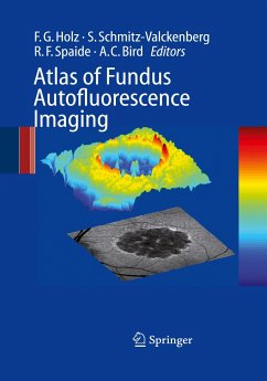
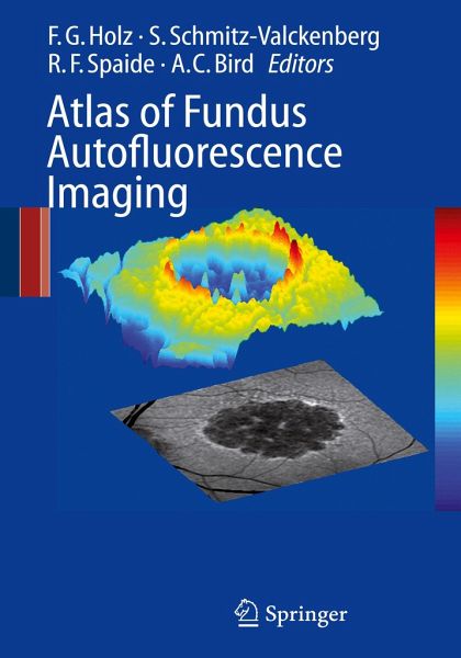
During recent years, FAF (Fundus autofluorescence) imaging has been shown to be useful in various retinal diseases with regard to diagnostics, documentation of changes, identification of disease progression, and monitoring of novel therapies. Hereby, FAF imaging gives additional information above and beyond conventional imaging tools.This unique atlas provides a comprehensive and up-to-date overview of FAF imaging in retinal diseases. It also compares FAF findings with other imaging techniques such asfundus photograph, fluorescein- and ICG angiography as well as optical coherence tomography.Ge...
During recent years, FAF (Fundus autofluorescence) imaging has been shown to be useful in various retinal diseases with regard to diagnostics, documentation of changes, identification of disease progression, and monitoring of novel therapies. Hereby, FAF imaging gives additional information above and beyond conventional imaging tools.
This unique atlas provides a comprehensive and up-to-date overview of FAF imaging in retinal diseases. It also compares FAF findings with other imaging techniques such as
fundus photograph, fluorescein- and ICG angiography as well as optical coherence tomography.
General ophthalmologists as well as retina specialists will find this a very useful guide which illustrates typical FAF characteristics of various retinal diseases.


