Bernard Koong
Atlas of Oral and Maxillofacial Radiology
Bernard Koong
Atlas of Oral and Maxillofacial Radiology
- Gebundenes Buch
- Merkliste
- Auf die Merkliste
- Bewerten Bewerten
- Teilen
- Produkt teilen
- Produkterinnerung
- Produkterinnerung
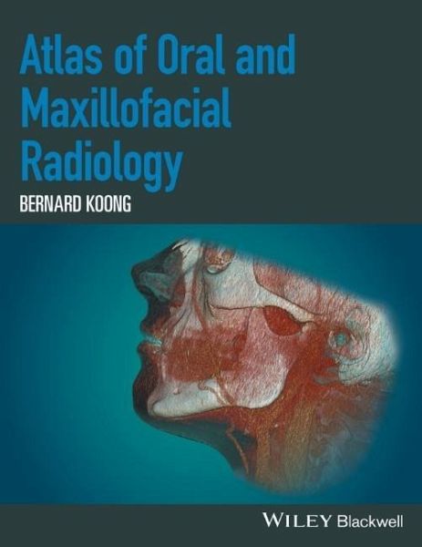
The Atlas of Oral and Maxillofacial Radiology presents an extensive case collection of both common and less common conditions of the jaws and teeth. Focusing on the essentials of radiologic interpretation, this is a go-to companion for clinicians in everyday practice who have radiologically identified a potential abnormality, as well as a comprehensive study guide for students at all levels of dentistry, surgery and radiology. _ Unique lesion-based problem solving chapter makes this an easy-to-use reference in a clinical setting _ Includes 2D intraoral radiography, the panoramic radiograph,…mehr
Andere Kunden interessierten sich auch für
- Michael A. KahnThe ADA Practical Guide to Soft Tissue Oral Disease107,99 €
- Oral and Maxillofacial Radiology215,99 €
- Salivary Gland Pathology337,99 €
- Diagnosing Dental and Orofacial Pain71,99 €
- Mark GreenwoodEssentials of Human Disease in Dentistry87,99 €
- Sexually Transmissible Oral Diseases145,99 €
- Eric CarlsonSalivary Gland Pathology233,99 €
-
-
-
The Atlas of Oral and Maxillofacial Radiology presents an extensive case collection of both common and less common conditions of the jaws and teeth. Focusing on the essentials of radiologic interpretation, this is a go-to companion for clinicians in everyday practice who have radiologically identified a potential abnormality, as well as a comprehensive study guide for students at all levels of dentistry, surgery and radiology.
_ Unique lesion-based problem solving chapter makes this an easy-to-use reference in a clinical setting
_ Includes 2D intraoral radiography, the panoramic radiograph, cone beam CT, multidetector CT and MRI
_ Multiple cases are presented in order to demonstrate the variation in the radiological appearances of conditions affecting the jaws and teeth
_ Special focus on conditions where diagnostic imaging may substantially contribute to diagnosis
Hinweis: Dieser Artikel kann nur an eine deutsche Lieferadresse ausgeliefert werden.
_ Unique lesion-based problem solving chapter makes this an easy-to-use reference in a clinical setting
_ Includes 2D intraoral radiography, the panoramic radiograph, cone beam CT, multidetector CT and MRI
_ Multiple cases are presented in order to demonstrate the variation in the radiological appearances of conditions affecting the jaws and teeth
_ Special focus on conditions where diagnostic imaging may substantially contribute to diagnosis
Hinweis: Dieser Artikel kann nur an eine deutsche Lieferadresse ausgeliefert werden.
Produktdetails
- Produktdetails
- Verlag: Wiley & Sons / Wiley-Blackwell
- Artikelnr. des Verlages: 1W118939640
- 1. Auflage
- Seitenzahl: 368
- Erscheinungstermin: 17. April 2017
- Englisch
- Abmessung: 281mm x 219mm x 22mm
- Gewicht: 1244g
- ISBN-13: 9781118939642
- ISBN-10: 1118939646
- Artikelnr.: 45075794
- Verlag: Wiley & Sons / Wiley-Blackwell
- Artikelnr. des Verlages: 1W118939640
- 1. Auflage
- Seitenzahl: 368
- Erscheinungstermin: 17. April 2017
- Englisch
- Abmessung: 281mm x 219mm x 22mm
- Gewicht: 1244g
- ISBN-13: 9781118939642
- ISBN-10: 1118939646
- Artikelnr.: 45075794
Bernard Koong is a highly experienced oral and maxillofacial radiologist practicing full time multimodality clinical radiology. He is a founding partner of Envision Medical Imaging, a multidisciplinary fully comprehensive private radiology group in Australia and also consults internationally. He has personally reported over 200,000 radiological studies involving a wide variety of imaging techniques. Having completed his specialist training in oral and maxillofacial radiology at the University of Toronto, Bernard now holds the position of Clinical Professor at the University of Western Australia, where he coordinates and delivers the oral and maxillofacial radiology lectures for the undergraduate and postgraduate courses. He also has a long history of providing oral and maxillofacial radiology courses for other universities as well as surgery and radiology programmes across Australasia. As an invited speaker, Bernard has presented more than 100 lectures to the dental and medical professions internationally, and is a member of the Editorial Board of Clinical Oral Implant Research.
List of Contributors, xi
Preface, xii
Acknowledgements, xiii
How to Use This Atlas, xiv
1 Problem Solving Diagrams, 1
1.1 Opaque and largely opaque conditions related to the jaws, 1
Common conditions, 1
Less common conditions, 1
1.2 Lucent lesions of the jaws, 2
Common conditions, 2
Less common conditions, 2
1.3 Mixed density lesions of the jaws, 3
Common conditions, 3
Less common conditions, 3
2 Radiological Anatomy, 4
2.1 The panoramic radiograph, 4
2.2 Identification of teeth - FDI (Fédération Dentaire Internationale) World Dental Federation notation, 8
2.3 Cone beam computed tomography, 11
Axial, 11
Sagittal, 18
Coronal, 22
3 Anomalies Related to the Teeth, 28
3.1 Supernumerary teeth, 28
3.2 Congenital absence, 30
3.3 Delayed and early development/eruption, 31
3.4 Ectopic development and eruption, 32
3.5 Impaction, 36
3.6 Macrodontia, 40
3.7 Microdontia, 41
3.8 Dilaceration, 42
3.9 Enamel pearl, 42
3.10 Talon cusp, 43
3.11 Dens invaginatus, 44
3.12 Dens evaginatus, 45
3.13 Taurodontism, 45
3.14 Fusion, 46
3.15 Gemination, 47
3.16 Concrescence, 47
3.17 Amelogenesis imperfecta, 48
3.18 Dentinogenesis imperfecta, 49
3.19 Dentin dysplasia, 50
3.20 Secondary and tertiary dentin, 51
3.21 Pulp stones, 52
3.22 Hypercementosis, 53
4 Conditions Related to Loss of Tooth Structure, 54
4.1 Caries, 54
Interproximal caries, 54
Pit and fissure caries, 54
Root caries, 55
4.2 Attrition, 59
4.3 Abrasion, 60
4.4 Erosion, 61
4.5 Internal resorption, 61
4.6 External resorption, 62
4.7 Fracture related to trauma, 63
5 Inflammatory Lesions of the Jaws, 64
5.1 Periapical inflammatory lesions, 64
Post-treatment appearances of periapical lesions, 65
Re-establishment of normal periapical structures, 65
Variant trabecular architecture, 65
Fibrous healing, 65
Periapical osseous prominence at the maxillary sinus base, 66
5.2 Periodontal inflammatory disease, 74
5.3 Pericoronitis, 83
5.4 Osteomyelitis of the jaws, 86
5.5 Dentoalveolar and jaw infections involving the adjacent soft tissues, 88
6 Osteoradionecrosis and Osteonecrosis of the Jaws, 92
6.1 Osteoradionecrosis of the jaws, 92
6.2 Osteonecrosis of the jaws, 96
7 Hamartomatous/Hyperplastic Bony Opacities and Prominences Involving the Jaws, 97
7.1 Torus palatinus, 97
7.2 Torus mandibularis, 98
7.3 Exostoses, 100
7.4 Bone island, 101
8 Cysts and Cyst-like Lesions Involving the Jaws, 108
Odontogenic cysts and cyst -like lesions, 108
8.1 Radicular cyst, 108
8.2 Residual cyst, 114
8.3 Dentigerous cyst, 115
8.4 Buccal bifurcation cyst, 122
8.5 Keratocystic odontogenic tumour, 124
8.6 Basal cell naevus syndrome, 127
8.7 Lateral periodontal cyst, 128
8.8 Glandular odontogenic cyst, 130
Non-odontogenic cysts and cyst -like lesions, 130
8.9 Simple bone cyst, 130
8.10 Nasopalatine duct cyst, 136
8.11 Nasolabial cyst, 138
9 Fibro-osseous Lesions of the Jaws, 140
9.1 Fibrous