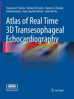
This atlas provides a comprehensive description of normal anatomy of the internal structures of the heart (natives valves, interatrial septum, left atrial appendage, left atrium etc..) as seen by this revolutionary ultrasound technique. Normal TEE cardiac structures are described and compared with the corresponding anatomical specimens focusing on the fundamental as well as the details of the cardiac structures, providing a detailed understanding of the anatomy that has not previously been possible with either real-time transthoracic echocardiography (TTE) or reconstructed 3D TEE imaging technology. The atlas contains a large number of challenging cardiac pathology cases observed in clinical settings and based of the combined experience of five outstanding institutions in Europe and United States. Each case is accompanied by a brief presentation and discussion of the value of the imaging modality to effective diagnosis.
Bitte wählen Sie Ihr Anliegen aus.
Rechnungen
Retourenschein anfordern
Bestellstatus
Storno

