Electron microscopy is frequently portrayed as a discipline that stands alone, separated from molecular biology, light microscopy, physiology, and biochemistry, among other disciplines. It is also presented as a technically demanding discipline operating largely in the sphere of "black boxes" and governed by many absolute laws of procedure. At the introductory level, this portrayal does the discipline and the student a disservice. The instrumentation we use is complex, but ultimately understandable and, more importantly, repairable. The procedures we employ for preparing tissues and cells are not totally understood, but enough information is available to allow investigators to make reasonable choices concerning the best techniques to apply to their parti cular problems. There are countless specialized techniques in the field of electron and light microscopy that require the acquisition of specialized knowledge, particularly for interpretation of results (electron tomography and energy dispersive spectroscopy immediately come to mind), but most laboratories possessing the equipment to effect these approaches have specialists to help the casual user. The advent of computer operated electron microscopes has also broadened access to these instruments, allowing users with little technical knowledge about electron microscope design to quickly become operators. This has been a welcome advance, because earlier instru ments required a level of knowledge about electron optics and vacuum systems to produce optimal photographs and to avoid "crashing" the instruments that typically made it difficult for beginners.
Hinweis: Dieser Artikel kann nur an eine deutsche Lieferadresse ausgeliefert werden.
Hinweis: Dieser Artikel kann nur an eine deutsche Lieferadresse ausgeliefert werden.
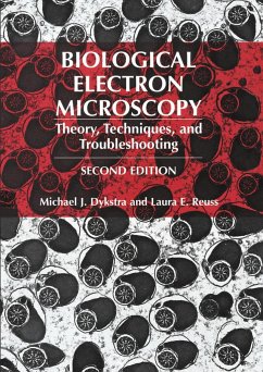
"In this second edition of his 1992 hardcover text and 1993 spiral-bound lab manual on Biological Electron Microscopy, Michael Dykstra has expended considerable effort to merge the two earlier volumes into a more readable and usable single volume and also to update them and add considerable new material. Happily, the result is a first rate, comprehensive book that will be useful for both teaching beginning students and as a reference book for experienced researchers. In all chapters, new materials have been added in the text and referenced at the chapter ends, and where appropriate, useful web sites have been indicated where additional information may be obtained. All of the excellent illustrative photographs from the first edition have been retained. As before, the chapters on microscope construction and operation are very well done and will be invaluable in teaching. Cautionary statements are made throughout about handling and disposal of the hazardous materials used in EM labs. Relevant journals, societies, and equipment suppliers are listed in appendices. In general, this single volume is a welcome addition to the literature available on biological microscopy. Its publication offers support for the idea that microscopy in all of its guises is still a dynamic and valuable tool for all biologists." (Henry C. Aldrich, Professor Emeritus of Microbiology and Cell Science, University of Florida, Gainesville)


