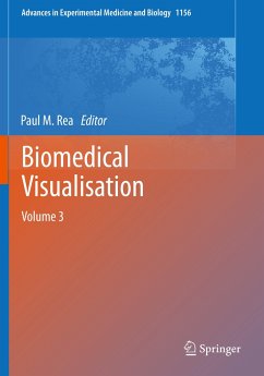
This edited book explores the use of technology to enable us to visualise the life sciences in a more meaningful and engaging way. It will enable those interested in visualisation techniques to gain a better understanding of the applications that can be used in visualisation, imaging and analysis, education, engagement and training.
The reader will be able to explore the utilisation of technologies from a number of fields to enable an engaging and meaningful visual representation of the biomedical sciences, with a focus in this volume related to anatomy, and clinically applied scenarios.
The first six chapters have an anatomical focus examining digital technologies and applications to enhance education. The first examines the history and development of ultrasound, applications in an educational setting, and as a point-of-care ultrasound at the bedside. The second chapter presents a transferable workflow methodology in creating an interactive educational and trainingpackageto enhance understanding of the circadian rhythm. The third chapter reviews tools and technologies, which can be used to enhance off-campus learning, and the current range of visualisation technologies like virtual, augmented and mixed reality systems. Chapter four discusses how scanning methodologies like CT imagery, can make stereoscopic models. The fifth chapter describes a novel way to reconstruct 3D anatomy from imaging datasets and how to build statistical 3D shape models, described in a clinical context and applied to diagnostic disease scoring. The sixth chapter looks at interactive visualisations of atlases in the creation of a virtual resource, for providing next generation interfaces.
The seventh and eight chapters discuss neurofeedback for mental health education and interactive visual data analysis (applied to irritable bowel disease) respectively.
The final two chapters examine current immersive technologies -virtual and augmented reality, with the last chapter detailing virtual reality in patients with dementia.
This book is accessible to a wide range of users from faculty and students, developers and computing experts, the wider public audience. It is hoped this will aid understanding of the variety of technologies which can be used to enhance understanding of clinical conditions using modern day methodologies.

