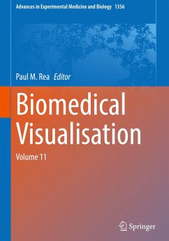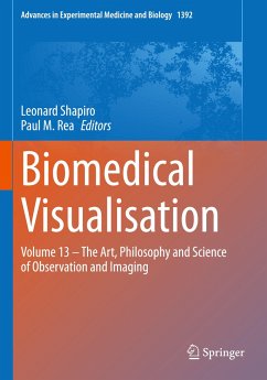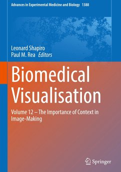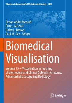
This edited book explores the use of technology to enable us to visualise the life sciences in a more meaningful and engaging way. It will enable those interested in visualisation techniques to gain a better understanding of the applications that can be used in visualisation, imaging and analysis, education, engagement and training. The reader will also be able to learn about the use of visualisation techniques and technologies for the historical and forensic settings. The chapters presented in this volume cover such a diverse range of topics, with something for everyone. We present here…mehr
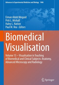
This edited book explores the use of technology to enable us to visualise the life sciences in a more meaningful and engaging way. It will enable those interested in visualisation techniques to gain a better understanding of the applications that can be used in visualisation, imaging and analysis, education, engagement and training. The reader will also be able to learn about the use of visualisation techniques and technologies for the historical and forensic settings.
The chapters presented in this volume cover such a diverse range of topics, with something for everyone. We present here chapters on 3D visualising novel stent grafts to aid treatment of aortic aneuryms; confocal microscopy constructed vascular models in patient education; 3D patient specific virtual reconstructions in surgery; virtual reality in upper limb rehabilitation in patients with multiple sclerosis and virtual clinical wards. In addition, we present chapters in artificial intelligence in ultrasoundguided regional anaesthesia; carpal tunnel release visualisation techniques; visualising for embryology education and artificial intelligence data on bone mechanics. Finally we conclude with chapters on visualising patient communication in a general practice setting; digital facial depictions of people from the past; instructor made cadaveric videos, novel cadaveric techniques for enhancing visualisation of the human body and finally interactive educational videos and screencasts.
This book explores the use of technologies from a range of fields to provide engaging and meaningful visual representations of the biomedical sciences. It is therefore an interesting read for researchers, developers and educators who want to learn how visualisation techniques can be used successfully for a variety of purposes, such as educating students or training staff, interacting with patients and biomedical procedures in general.
Produktdetails
Produktdetails
Advances in Experimental Medicine and Biology 1356
Die Herstellerinformationen sind derzeit nicht verfügbar.
Autorenporträt
Paul M. Rea is a Professor of Digital and Anatomical Education at the University of Glasgow. He is qualified with a medical degree (MBChB), a MSc (by research) in craniofacial anatomy/surgery, a PhD in neuroscience, the Diploma in Forensic Medical Science (DipFMS), and an MEd with Merit (Learning and Teaching in Higher Education). He is an elected Fellow of the Royal Society for the encouragement of Arts, Manufactures and Commerce (FRSA), elected Fellow of the Royal Society of Biology (FRSB), Senior Fellow of the Higher Education Academy, professional member of the Institute of Medical Illustrators (MIMI) and a registered medical illustrator with the Academy for Healthcare Science. Paul has published widely and presented at many national and international meetings, including invited talks. He sits on the Executive Editorial Committee for the Journal of Visual Communication in Medicine, is Associate Editor for the European Journal of Anatomy and reviews for 25 different journals/publishers. He is the Public Engagement and Outreach lead for anatomy coordinating collaborative projects with the Glasgow Science Centre, NHS and Royal College of Physicians and Surgeons of Glasgow. Paul is also a STEM ambassador and has visited numerous schools to undertake outreach work. His research involves a long-standing strategic partnership with the School of Simulation and Visualisation The Glasgow School of Art. This has led to multi-million pound investment in creating world leading 3D digital datasets to be used in undergraduate and postgraduate teaching to enhance learning and assessment. This successful collaboration resulted in the creation of the worlds first taught MSc Medical Visualisation and Human Anatomy combining anatomy and digital technologies. The Institute of Medical Illustrators also accredits it. It has created college-wide, industry, multi-institutional and NHS research linked projects for students. Paul is the Programme Director for this degree.
Inhaltsangabe
Chapter 1. Creating Interactive Three-Dimensional Applications to Visualise Novel Stent Grafts That Aid in the Treatment of Aortic Aneurysms.- Chapter 2. Using Confocal Microscopy to Generate an Accurate Vascular Model for Use in Patient Education Animation.- Chapter 3. Methods and applications of 3D Patient-Specific Virtual Reconstructions in Surgery.- Chapter 4. Proof of Concept for the Use of Immersive Virtual Reality in Upper Limb Rehabilitation of Multiple Sclerosis Patients.- Chapter 5. Virtual Wards: A Rapid Adaptation to Clinical Attachments in MBChB During the COVID-19 Pandemic.- Chapter 6. Artificial Intelligence: Innovation to Assist in the Identification of Sono-anatomy for Ultrasound-Guided Regional Anaesthesia.- Chapter 7. A Systematic Review of Randomised Control Trials Evaluating the Efficacy and Safety of Open and Endoscopic Carpal Tunnel Release.- Chapter 8. Exploring Visualisation for Embryology Education: A Twenty-First-Century Perspective.- Chapter 9. How Artificial Intelligence and Machine Learning Is Assisting Us to Extract Meaning from Data on Bone Mechanics?.- Chapter 10. Visual Communication and Creative Processes Within the Primary Care Consultation.- Chapter 11. Digital 2D, 2.5D and 3D Methods for Adding Photo-Realistic Textures to 3D Facial Depictions of People from the Past.- Chapter 12. Teaching with Cadavers Outside of the Dissection Room Using Cadaveric Videos.- Chapter 13. A Novel Cadaveric Embalming Technique for Enhancing Visualisation of Human Anatomy.- Chapter 14. Assessing the Impact of Interactive Educational Videos and Screencasts Within Pre-clinical Microanatomy and Medical Physiology Teaching.
Es gelten unsere Allgemeinen Geschäftsbedingungen: www.buecher.de/agb
Impressum
www.buecher.de ist ein Internetauftritt der buecher.de internetstores GmbH
Geschäftsführung: Monica Sawhney | Roland Kölbl | Günter Hilger
Sitz der Gesellschaft: Batheyer Straße 115 - 117, 58099 Hagen
Postanschrift: Bürgermeister-Wegele-Str. 12, 86167 Augsburg
Amtsgericht Hagen HRB 13257
Steuernummer: 321/5800/1497
USt-IdNr: DE450055826