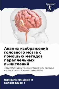
BRAIN IMAGE ANALYSIS USING PARALLEL COMPUTING TECHNIQUES
Medical Image Processing with High Performance Computing
Versandkostenfrei!
Versandfertig in 6-10 Tagen
53,99 €
inkl. MwSt.

PAYBACK Punkte
27 °P sammeln!
Medical images of human body concentrate on capturing of images from organs for both diagnostic and therapeutic purposes. Magnetic resonance imaging (MRI) is a non-invasive diagnostic test that takes detailed images of the soft tissues of the body. The technologies and automatic methods are more useful in several medical imaging applications such as volumetric analysis, three dimensional (3D) visualization, surgical planning, and simulations for detecting brain related diseases such as brain Tumors, multiple Sclerosis, Schizophrenia, Epilepsy, Parkinson's disease, Alzheimer's disease, and othe...
Medical images of human body concentrate on capturing of images from organs for both diagnostic and therapeutic purposes. Magnetic resonance imaging (MRI) is a non-invasive diagnostic test that takes detailed images of the soft tissues of the body. The technologies and automatic methods are more useful in several medical imaging applications such as volumetric analysis, three dimensional (3D) visualization, surgical planning, and simulations for detecting brain related diseases such as brain Tumors, multiple Sclerosis, Schizophrenia, Epilepsy, Parkinson's disease, Alzheimer's disease, and other pathologies. The automatic methods for brain tissue segmentation, tumor slices detection, and tumor segmentation with substructures are always on demand in the field of medical image processing due to limited human resources, time constraints, accuracy, and artefacts. In this book, five automatic methods are developed and presented with three parallel models for brain tumor analysis in thechapter 4 - 6.


