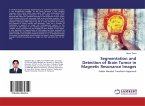
An efficient technique is proposed for the precise segmentation of normal and pathological tissues in the MRI brain images. The proposed segmentation technique initially performs classification process by utilizing K-means clustering. This proposes a Herpes Simplex Virus approach for classification of brain magnetic resonance images (MRI) based on colour converted K-means clustering segmentation algorithm. Segmentation of images holds an important position in the area of image processing. It becomes more important while typically dealing with medical images. A well known segmentation problem within MRI is the task of labeling voxels according to their tissue type which include White Matter, Grey Matter, Cerebrospinal Fluid and sometimes pathological tissues like tumour etc. This thesis describes an efficient method for automatic brain tumour segmentation.
Hinweis: Dieser Artikel kann nur an eine deutsche Lieferadresse ausgeliefert werden.
Hinweis: Dieser Artikel kann nur an eine deutsche Lieferadresse ausgeliefert werden.


