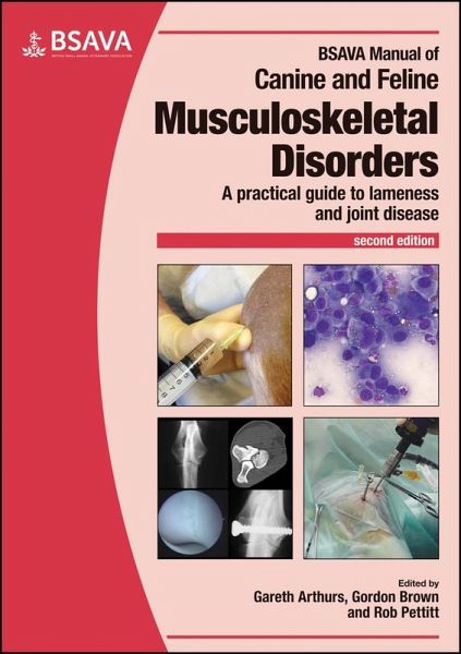
BSAVA Manual of Canine and Feline Musculoskeletal Disorders
Herausgeber: Arthurs, Gareth; Pettit, Robert; Brown, Gordon D. A.
BSAVA Manual of Canine and Feline Musculoskeletal Disorders
Herausgeber: Arthurs, Gareth; Pettit, Robert; Brown, Gordon D. A.
- Broschiertes Buch
- Merkliste
- Auf die Merkliste
- Bewerten Bewerten
- Teilen
- Produkt teilen
- Produkterinnerung
- Produkterinnerung
This new edition builds upon the success of the first edition of the BSAVA Manual of Canine and Feline Musculoskeletal Disorders, as well as its predecessor the BSAVA Manual of Small Animal Arthrology. The chapters have been extensively revised and updated to reflect the advances in the field over the past 10 years. The manual is divided into four main sections: * Lameness investigation * Musculoskeletal disorders * Principles of orthopaedic surgery * Management of specific disorders, ordered by joint.
Andere Kunden interessierten sich auch für
- BSAVA Manual of Canine and Feline Gastroenterology123,99 €
- BSAVA Manual of Canine and Feline Thoracic Imaging122,99 €
- BSAVA Manual of Canine and Feline Endoscopy and Endosurgery123,99 €
- BSAVA Manual of Canine and Feline Endocrinology117,99 €
- BSAVA Manual of Canine and Feline Nephrology and Urology130,99 €
- BSAVA Pocketbook for Vets41,99 €
- BSAVA Guide to Pain Management in Small Animal Practice65,99 €
-
-
-
This new edition builds upon the success of the first edition of the BSAVA Manual of Canine and Feline Musculoskeletal Disorders, as well as its predecessor the BSAVA Manual of Small Animal Arthrology. The chapters have been extensively revised and updated to reflect the advances in the field over the past 10 years. The manual is divided into four main sections: * Lameness investigation * Musculoskeletal disorders * Principles of orthopaedic surgery * Management of specific disorders, ordered by joint.
Hinweis: Dieser Artikel kann nur an eine deutsche Lieferadresse ausgeliefert werden.
Produktdetails
- Produktdetails
- BSAVA British Small Animal Veterinary Association
- Verlag: British Small Animal Veterinary Association
- 2 ed
- Seitenzahl: 504
- Erscheinungstermin: 21. Dezember 2018
- Englisch
- Abmessung: 296mm x 209mm x 27mm
- Gewicht: 1638g
- ISBN-13: 9781905319695
- ISBN-10: 190531969X
- Artikelnr.: 54828991
- Herstellerkennzeichnung
- Libri GmbH
- Europaallee 1
- 36244 Bad Hersfeld
- 06621 890
Gareth Arthurs, PGCertMedEd, MA, VetMB, CertVR, CertSAS, DSAS(Orth), FHEA, FRCVS, RCVS Recognised Specialist in Small Animal Surgery (Orthopaedics), graduated from Cambridge University in 1996 and spent time in mixed and first opinion practice before concentrating on small animal orthopaedics. He has been a UK RCVS Diplomate and recognised specialist since 2007 and has worked at a number of tertiary referral centres. He is senior vice chairman of the British Veterinary Orthopaedic Association, honorary associate professor in small animal orthopaedic surgery at the University of Nottingham, an examiner for the Royal College of Veterinary Surgeons and a reviewer for a number of international journals. Gordon Brown, BVM&S, Cert SAO, DSAS (Orth), MRCVS, RCVS Diplomate in Small Animal Surgery (Orthopaedics), graduated from the 'Dick Vet' in 1984. He has over 20 years experience of small animal orthopaedic referral work, gaining his RCVS Diploma from practice in 2017. He is co-founder and currently referral director of the Grove Referrals in Norfolk. He was involved as series editor on the first edition of this manual. He is a member of the AOVET national faculty and is currently chair of the BVOA. Rob Pettitt, BVSc, PGCertLTHE, DSAS (Orth), FHEA, MRCVS, RCVS Specialist in Small Animal Surgery (Orthopaedics), graduated from the University of Liverpool in 2002 and spent three years in general practice before returning to Liverpool as a Clinician Teacher in Small Animal Orthopaedics. Rob passed the RCVS Diploma in Small Animal Orthopaedics exam in 2010 and is a current examiner for the final years of the examination. Rob is a current member of the AOVET national faculty and a Senior Lecturer at the University of Liverpool.
List of contributors
Foreword
Preface
Section 1: Lameness investigation
1 Normal locomotion
Neil Burton and Gordon Brown
2 Lameness examination
Harry Scott
3 Imaging
Thomas W. Maddox
4 Sampling and laboratory investigation
Emma Scurrell and Gareth Arthurs
Section 2: Musculoskeletal disorders
5 The normal joint
Richard Barrett Jolley and Rebecca Lewis
6 Arthritis
Ralph Abercromby, John Innes and Dylan Clements
7 Diseases and disorders of bone
Gareth Arthurs and Sorrel Langley-Hobbs
8 Disturbances of growth and bone development
Gareth Arthurs and Sorrel Langley-Hobbs
9 Muscle, tendon and ligament injuries
Angus Anderson
10 Diseases of muscle and the neuromuscular junction
G. Diane Shelton
11 Tumours of the musculoskeletal system
Nicholas J. Bacon
12 Neurological causes of lameness
Sebastien Behr and Simona Tiziana Radaelli
Section 3: Principles of orthopaedic surgery
13 Principles of orthopaedic surgery
Rob Pettitt
14 Surgical instruments and implants
John Lapish and David Strong
15 Arthroscopy
Bernadette Van Ryssen and John Lapish
16 Complications of orthopaedic surgery
Simon Roch
17 Postoperative management and rehabilitation
Briony Alderson, Rob Pettitt and Louise Dale
Section 4: Management of specific disorders, ordered by individual joints
18 The shoulder
Steve Butterworth and Rob Pettitt
Imaging Techniques: Thomas W. Maddox
19 The elbow
Neil Burton
Imaging Techniques: Thomas W. Maddox
20 The carpus
Stephen Clarke and Jonathan Pink
Imaging Techniques: Thomas W. Maddox
21 The distal limb
Mike Guilliard
Imaging Techniques: Thomas W. Maddox
22 The hip
Toby J. Gemmill and Bill Oxley
Imaging Techniques: Thomas W. Maddox
23 The Stifle Mike Farrell
Imaging Techniques: Thomas W. Maddox
24 The tarsus
Rob Pettitt, Gareth Arthurs, Brian Beale Don Hulse
Imaging Techniques: Thomas W. Maddox
25 The temporomandibular joint
Andy Moores
Imaging Techniques: Thomas W. Maddox
Index