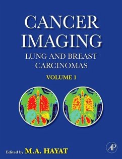
With cancer-related deaths projected to rise to 10.3 million people by 2020, the need to prevent, diagnose, and cure cancer is greater than ever. This book presents readers with the most up-to-date imaging instrumentation, general and diagnostic applications for various cancers, with an emphasis on lung and breast carcinomas--the two major worldwide malignancy types. This book discusses the various imaging techniques used to locate and diagnose tumors, including ultrasound, X-ray, color Doppler sonography, PET, CT, PET/CT, MRI, SPECT, diffusion tensor imaging, dynamic infrared imaging, and magnetic resonance spectroscopy. It also details strategies for imaging cancer, emphasizing the importance of the use of this technology for clinical diagnosis. Imaging techniques that predict the malignant potential of cancers, response to chemotherapy and other treatments, recurrence, and prognosis are also detailed.
. Concentrates on the application of imaging technology to the diagnosis and prognosis of lung and breast carcinomas, the two major worldwide malignancies
. Addresses the relationship between radiation dose and image quality
. Discusses the role of molecular imaging in identifying changes for the emergence and progression of cancer at the cellular and/or molecular levels
Hinweis: Dieser Artikel kann nur an eine deutsche Lieferadresse ausgeliefert werden.
"Professor Hayat has assembled a distinguished group of scientists and clinicians from around the world and has provided a very timely discussion of functional imaging for the diagnosis and treatment of solid organ cancers. Dr. Hayat's introduction and discussion of imaging techniques are comprehensive. He has clearly outlined the roles of functional imaging in defining the nature of primary organ cancers and their metastases.
-- Akhouri A. Sinha, Professor of Genetics, Cell Biology, and Development, University of Minnesota; Senior Research Scientists, Veterans Affairs Medical Center, Minneapolis, MN
-- Akhouri A. Sinha, Professor of Genetics, Cell Biology, and Development, University of Minnesota; Senior Research Scientists, Veterans Affairs Medical Center, Minneapolis, MN

