- Gebundenes Buch
- Merkliste
- Auf die Merkliste
- Bewerten Bewerten
- Teilen
- Produkt teilen
- Produkterinnerung
- Produkterinnerung
This book introducing all researchers and students who are interested in pursuing their research in the field of application of carbon dots in health care especially as a theranostic agent. It focuses on the fundamental understanding along with the applications of this unique fluorescent nanobiomachine, the Carbon Dot. The book begins with the explanation that carbon dots fall between the usual daily macro or bulk physics and the quantum mechanics and covers their unique properties like quantum mechanics and quantum confinement. It then encompasses the domain of various physical, chemical and…mehr
Andere Kunden interessierten sich auch für
- Carbon Nanofibers252,99 €
- Industrial Carbon and Graphite Materials306,99 €
- Functionalized Carbon Nanotubes for Biomedical Applications239,99 €
- Emerging Carbon-Based Nanocomposites for Environmental Applications247,99 €
- Enhanced Carbon-Based Materials and Their Applications124,99 €
- Ashutosh TiwariAdvanced Theranostic Materials233,99 €
- Graphene209,99 €
-
-
-
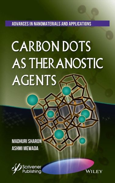
This book introducing all researchers and students who are interested in pursuing their research in the field of application of carbon dots in health care especially as a theranostic agent. It focuses on the fundamental understanding along with the applications of this unique fluorescent nanobiomachine, the Carbon Dot. The book begins with the explanation that carbon dots fall between the usual daily macro or bulk physics and the quantum mechanics and covers their unique properties like quantum mechanics and quantum confinement. It then encompasses the domain of various physical, chemical and biological methods that efficiently synthesizes the carbon dots and their desired properties. The basic characterization techniques used for carbon dots is also covered in this book. Conjugation of carbon dots with different moieties is another aspect that enhances its applications, hence this is highlighted too. The final attributes of this book is that how to maneuver the carbon dots for their use in targeted drug delivery with emphasis on cancer and neurodegenerative disease as well as cellular imaging and diagnostics. One of the unique features of this book is that it has looked into the use of carbon dots to act as a nanofertilizer, as a drug/antibiotic delivery vehicle to diseased plants through foliar application.
Hinweis: Dieser Artikel kann nur an eine deutsche Lieferadresse ausgeliefert werden.
Produktdetails
- Produktdetails
- Verlag: Wiley & Sons / Wiley-Scrivener
- Artikelnr. des Verlages: 1W119460100
- 1. Auflage
- Seitenzahl: 288
- Erscheinungstermin: 5. September 2018
- Englisch
- Abmessung: 235mm x 157mm x 20mm
- Gewicht: 570g
- ISBN-13: 9781119460107
- ISBN-10: 1119460107
- Artikelnr.: 50443259
- Verlag: Wiley & Sons / Wiley-Scrivener
- Artikelnr. des Verlages: 1W119460100
- 1. Auflage
- Seitenzahl: 288
- Erscheinungstermin: 5. September 2018
- Englisch
- Abmessung: 235mm x 157mm x 20mm
- Gewicht: 570g
- ISBN-13: 9781119460107
- ISBN-10: 1119460107
- Artikelnr.: 50443259
Madhuri Sharon, (Retd Director Reliance Industries LTD) PhD from Leicester University UK, Post-Doctoral Research from Bolton Institute of Technology U.K., is currently Director of NSN Research Centre for Nanotechnology & Bionanotechnology and Managing Director of Monad Nanotech as well as Adjunct-Professor University of Mumbai & Professor-Emeritus JJT University. She has published more than 130 papers, 4 books and 11 patents. Her research focuses on the synthesis, biosynthesis and application of various nanomaterials (graphene oxide, carbon dots, carbon nanomaterials and nanometals, SPIONS) in drug-delivery. Ashmi Mewada holds a PhD degree from Nagoya Institute of Technology, Japan. She is currently an Assistant Professor in the Department of Nanotechnology at Walchand College of Arts and Science, Solapur. She has more than 30 publications and works extensively in the field of carbon dots.
Preface
1. Carbon Dots: Discovery, Synthesis and Characterization 1
1.1. Background 1
1.2. Introduction to QD 2
1.2.1. What is Quantum Mechanics? 4
1.2.2. Quantum Confinement 7
1.2.3. Discovery and History of Carbon Dots 8
1.3. Carbon QD and Graphene QD 9
1.4. Various Methods of Synthesis of Carbon Dots 10
1.4.1. Electrochemical Methods 11
1.4.2. Combustion and Thermal Oxidation Method 13
1.4.3. Hydrothermal Oxidation Method 15
1.4.4. Solvothermal Method 18
1.4.5. Laser Ablation of Graphite 18
1.4.6. Pulsed Laser Irradiation of Carbon Source 20
1.4.7. Arc Discharge Method 20
1.4.8. Plasma Treatment 21
1.4.9. Opening of Fullerene Cage 22
1.4.10. Ultrasonication Method 22
1.4.11. Microwave-Assisted Method 23
1.4.12. Chemical Methods 26
1.4.13. Supported Synthetic Procedure 26
1.4.14. Biogenic Method 28
1.5. Characterization of Carbon Dots 31
1.5.1. Microscopic Methods 32
1.5.1.1. SEM and TEM Characterization 32
1.5.1.2. AFM and STM Characterization 34
1.5.2. Spectroscopic Methods 35
1.5.2.1. UV-Vis Spectroscopy and its Application for Band Gap Determination
37
1.5.2.2. Fluorescence Spectrometry 37
1.5.2.3. Fourier Transform Infrared (FTIR) Spectroscopy 38
1.5.2.4. X-Ray Diffraction (XRD) Analysis 40
1.5.2.5. X-Ray Photoelectron Spectroscopy (XPS) 41
1.5.2.6. Dynamic Light Scattering/Photon Correlation Spectroscopy (DLS/PCS)
41
1.5.2.7. Dual Polarization Interferometry (DPI) 42
1.5.2.8. Raman Spectroscopy 43
1.5.2.9. Nuclear Magnetic Resonance (NMR) Spectroscopy 44
1.6. Summary 45
2. Properties of Carbon Dots 47
2.1. Introduction 47
2.2. Optical Properties 49
2.2.1. Absorbance 51
2.2.2. Photo-Induced Electron Transfer (PET) with CDs 52
2.2.3. Fluorescence/Photoluminescence (PL) 53
2.2.3.1. Multiphoton Excitation 60
2.2.3.2. Upconversion Photoluminescence 61
2.2.3.3. Lack of Blinking 63
2.2.3.4. Resistance to Photobleaching 64
2.2.4. Photocatalytic Property 65
2.3. Chemically Inert 67
2.4. Easy Functionalization 67
2.5. Water Solubility 68
2.6. Low Toxicity 68
2.7. Biocompatibility 69
2.8. Summary 70
3. Carbon Dots and Conjugates 71
3.1. Introduction 71
3.2. Why Conjugation of Carbon Dots? 74
3.3. Types of Carbon Dot Conjugates and Their Applications 76
3.3.1. Biogenic Compounds Conjugated with Carbon Dots 77
3.3.1.1. CDs Conjugated with Proteins/Peptides 78
3.3.1.2. CD Conjugates of Amino: Carboxylic Acid Ratio 80
3.3.1.3. CDs Conjugated with DNA 80
3.3.1.4. CDs Conjugated with RNase and SiRNA 84
3.3.1.5. CDs Conjugated with Lipid 86
3.3.1.6. CDs Conjugated with Folic Acid 86
3.3.1.7. CDs Conjugated with Chitosan 88
3.3.1.8. CDs Conjugated with Digitonin 89
3.3.2. Inorganic Heteroatoms Conjugated with CDs 90
3.3.2.1. CDs Conjugated with Gold Nanoparticles 91
3.3.2.2. CDs Conjugated with Silica 92
3.3.2.3. CDs Conjugated with ZnO 94
3.3.2.4. CDs Conjugated with CdS 95
3.3.2.5. CDs Conjugated with Strontium Oxide 96
3.3.2.6. CDs Conjugated with Gadolinium(III) 97
3.3.2.7. CDs Conjugated with Europium 97
3.3.2.8. CDs Conjugated/Doped with Nitrogen, Sulphur, Phosphorus and Boron
99
3.3.3. Carbon Dots Conjugated with Organic Material 100
3.3.3.1. PEG (Polyethylene Glycol) 101
3.3.3.2. CDs Conjugated with PEI (Polyethylenimin) or Polyaziridine 102
3.3.3.3. CDs Conjugated with ¿-Cyclodextrin 105
3.3.3.4. CDs Conjugated with Cysteamine 106
3.3.3.5. CDs Conjugated with Dihydrolipoic Acid 106
3.3.3.6. CDs Conjugated with Polyamidoamine (PAMAM) Dendrimers 107
3.3.3.7. CDs Covalently Conjugated with Rhodamine B Dyes 108
3.3.3.8. CDs Conjugated with Fe-Aminoclay (FeAC) 109
3.3.3.9. CDs Conjugated with MWCNT 109
3.3.4. CDs Conjugated with Antibiotics 110
3.3.4.1. CDs Conjugated with Ciprofloxacin 111
3.3.4.2. CDs Conjugated with Tetracycline 114
3.3.4.3. CDs Conjugated with Vancomycin 114
3.3.4.4. CDs Conjugated with Ampicillin 115
3.3.4.5. CDs Conjugated with Streptomycin 116
3.3.5. CDs Conjugated with Anti-Neurodegenerative Drugs for Delivery to
Central Nervous System 118
3.3.5.1. CDs Conjugated with Haloperidol 119
3.3.5.2. CDs Conjugated with Transferrin 124
3.3.5.3. CDs Conjugated with Curcumin 125
3.3.6. CDs Conjugated with Anticancer Drugs 128
3.3.6.1. CDs Conjugated with Doxorubicin 128
3.3.6.2. CDs Conjugated with Cisplatin 130
3.4. Summary 132
4. CD as Drug Delivery Vehicle 133
4.1. Introduction 133
4.2. Considerations in Using CD as Drug Delivery Vehicle 136
4.3. Designs of CD-Based Drug Delivery System 137
4.3.1. Designing for Water-Insoluble Drugs 138
4.3.2. Designing for Targeting Tumor Location 138
4.3.3. Designing a Theranostic Nanomedicine 139
4.3.4. Designing a Photoresponsive Nzzano Drug Delivery System 139
4.3.5. Designing for Gene Delivery 140
4.3.6. Designing for Antibiotics Delivery 141
4.4. Carbon Dots for Delivery of Anti-Cancer Drug 142
4.4.1. A Brief Introduction to Cancer 143
4.4.2. Necessity of Drug Targeting in Cancer Therapy 144
4.4.3. Targeting Angiogenesis with CD 144
4.4.4. Various CD Conjugates for Delivering Anti-Cancer Drug 145
4.4.5. CD for pH-Dependent Drug Release 146
4.4.6. CD for Drug Delivery to Renal Cancer 147
4.4.7. CD for Drug Delivery to Lung Cancer 148
4.4.8. CD for Drug Delivery to Breast Cancer 149
4.5. CD for Drug Delivery to Neurodegenerative Disease 150
4.6. CD for Gene Therapy 151
4.7. CD to Monitor Delivery of SiRNA 152
4.8. Challenges in Using CD as Drug Delivery Vehicle 152
4.8.1. Prevention of Drug from Biological Degradation 153
4.8.2. Effective Targeting 154
4.8.3. Patient Compliance 155
4.8.4. Cost Effectiveness 155
4.9. Suitability of CD-Conjugated Drugs 156
4.9.1. For Oral Drug Delivery 156
4.9.2. By Inhalation 156
4.9.3. As Transdermal Drug Delivery 157
4.9.4. As Injection 157
4.10. Summary 157
5. Carbon Dots for Cell Imaging and Diagnostics 159
5.1. Introduction 159
5.2. Bioimaging 162
5.2.1. Bioimaging of Cancerous Cells 166
5.2.1.1. HeLa Cells 168
5.2.1.2. Human Breast Cancer MCF-7 Cells and Human Breast Tumor Cells
MDA-MB-468 170
5.2.1.3. B16F11 and HEK293 Cells 171
5.2.1.4. Ehrlich Ascites Carcinoma (EAC) Cells 173
5.2.1.5. Human U87 Cell 173
5.2.1.6. MGC-803 Human Gastric Cancer Cells 174
5.2.1.7. A549 Adenocarcinomic Human Alveolar (Lung) Basal Epithelial Cells
175
5.2.1.8. Human Hepatocellular Carcinoma Cells 175
5.2.1.9. Kidney Proximal Tubule Cell Line(LLC-PK1) 176
5.2.1.10. C6 Glioma Cells 177
5.2.2. Bioimaging of Nucleus 178
5.2.3. Bioimaging of Virus 180
5.2.4. Bioimaging of Bacteria 181
5.2.5. Bioimaging of Drosophila melanogaster 183
5.3. CDs as Sensor 184
5.3.1. Intracellular Detection of Ions 185
5.3.1.1. Detection of Ag Ions 185
5.3.1.2. CD for Detection of Cu Ion 187
5.3.1.3. Detection of Fe3+ Ions 190
5.3.1.4. Detection of Hg2+ Ions 191
5.3.2. Detection of Small Molecules 192
5.3.2.1. Detection of Nitric Oxide 193
5.3.2.2. Detection of Phosphate 193
5.3.2.3. Detection of Reactive Oxygen Species 194
5.3.2.4. Detection of H2S 194
5.3.2.5. Detection of TNT 195
5.3.2.6. Detection of Hydroquinone 195
5.3.2.7. Detection of Surfactant 196
5.3.2.8. Detection of Humidity 197
5.3.3. Detection of Biological pH Value 198
5.3.4. Detection of Nucleic Acid 200
5.3.5. Detection of Vitamins 201
5.3.6. Detection of Protein and Enzymes 202
5.3.7. Detection of Glucose 202
5.3.8. Detection of Cancerous Cells 203
5.3.9. Detection of Dopamine Neurotransmitter 204
5.4. Concluding Remarks 206
6. Suitability of Carbon Dots as Payload for Plants 209
6.1. Introduction 209
6.2. Suitability of Carbon Dots as a Payload for Plant 211
6.2.1. Cytotoxicity of Carbon Dots 212
6.2.2. Carbon Dots and Plant Growth 213
6.2.2.1. Wheat 215
6.2.2.2. Green Beans 215
6.2.3. Is Cell Wall a Barrier for Carbon Dot Internalization? 216
6.2.4. Other Possible Routes for Entry of Carbon Dots 219
6.3. Carbon Dots and Plant Fertilizer 221
6.3.1. Nitrogen 222
6.3.2. Phosphorus 224
6.3.3. Potassium 225
6.3.4. Micronutrients 226
6.4. Need for Sensor to Detect 227
6.4.1. Plant Nutrient Status 228
6.4.2. Water 229
6.4.3. Pathological Status of Plant 231
6.4.4. Residual Pesticide, Herbicide or Insecticide in Plants 232
6.5. Carbon Dots and Foliar Application of Drugs on Plants 233
6.5.1. Cuticular Layer 233
6.5.2. Cell Wall 235
6.5.3. Plasma Membrane 235
6.6. Carbon Dots as Trojan Horse to Penetrate Foliar Surface for
Therapeutic Molecule Delivery 236
6.6.1. CD for Delivery of Antibiotics to Plants 237
6.7. Concluding Remarks 240
References 241