Audrey Paterson, Mike Thornton, Peter Carter
Chesneys Equipment for Student
Herausgeber: Carter, P H; Pirrie, J R; Milne, A.; Hyatt, A P; Thornton, M L; Paterson, A M
Audrey Paterson, Mike Thornton, Peter Carter
Chesneys Equipment for Student
Herausgeber: Carter, P H; Pirrie, J R; Milne, A.; Hyatt, A P; Thornton, M L; Paterson, A M
- Gebundenes Buch
- Merkliste
- Auf die Merkliste
- Bewerten Bewerten
- Teilen
- Produkt teilen
- Produkterinnerung
- Produkterinnerung
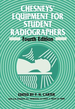
The new edition of this established text has been thoroughly revised and updated. It is divided into six parts. The first two parts cover the X-ray tube and X-ray generators. Part three looks at general, multipurpose radiographic equipment. Part four considers fluroscopic equipment, and the remaining two parts provide accounts of more specialized radiographic equipment and computer-based imaging modalities.
Andere Kunden interessierten sich auch für
![Chesneys' Radiographic Imaging Chesneys' Radiographic Imaging]() D. N. ChesneyChesneys' Radiographic Imaging105,99 €
D. N. ChesneyChesneys' Radiographic Imaging105,99 €![Index of Medical Imaging Index of Medical Imaging]() Jonathan McConnellIndex of Medical Imaging45,99 €
Jonathan McConnellIndex of Medical Imaging45,99 €![Essential Radiology Essential Radiology]() Richard B. GundermanEssential Radiology50,99 €
Richard B. GundermanEssential Radiology50,99 €![The Hands-On Guide to Imaging The Hands-On Guide to Imaging]() David C. HowlettThe Hands-On Guide to Imaging43,99 €
David C. HowlettThe Hands-On Guide to Imaging43,99 €![Radcases Plus Q&A Neuro Imaging Radcases Plus Q&A Neuro Imaging]() Radcases Plus Q&A Neuro Imaging49,99 €
Radcases Plus Q&A Neuro Imaging49,99 €![Radcases Ultrasound Imaging Radcases Ultrasound Imaging]() Nami R. AzarRadcases Ultrasound Imaging46,99 €
Nami R. AzarRadcases Ultrasound Imaging46,99 €![Elastography Elastography]() Richard G. BarrElastography87,99 €
Richard G. BarrElastography87,99 €-
-
-
The new edition of this established text has been thoroughly revised and updated. It is divided into six parts. The first two parts cover the X-ray tube and X-ray generators. Part three looks at general, multipurpose radiographic equipment. Part four considers fluroscopic equipment, and the remaining two parts provide accounts of more specialized radiographic equipment and computer-based imaging modalities.
Produktdetails
- Produktdetails
- Verlag: Blackwell Publishers
- 4th Edition
- Seitenzahl: 336
- Erscheinungstermin: 11. Mai 1994
- Englisch
- Abmessung: 244mm x 170mm x 18mm
- Gewicht: 581g
- ISBN-13: 9780632027248
- ISBN-10: 063202724X
- Artikelnr.: 13632088
- Herstellerkennzeichnung
- Libri GmbH
- Europaallee 1
- 36244 Bad Hersfeld
- gpsr@libri.de
P. H. Carter and A. M. Paterson are the authors of Chesneys' Equipment for Student Radiographers, 4th Edition, published by Wiley.
Part 1 The X-Ray Tube - Chapter 1 The X-Ray Tube: X-ray production
Electrical and radiation safety
Focal spot size
The problem of heat
X-ray tube construction and operation
Care of the X-ray tube
Follow-up practical
Part 2 X-Ray Generators - Chapter 2 Control of the X-Ray Tube Kilovoltage: Introduction
Voltage transformation
The high tension primary circuit
The need for rectification
Shortcomings of a pulsating X-ray supply
High tension cables
Kilovoltage compensation
Measuring kilovoltage
Follow-up practical
Chapter 3 Control of X-Ray Tube Current: Introduction
The need for accuracy
Tube filament circuitry
Falling load generators
Tube current measurement and display
Follow-up practical
Chapter 4 Exposure Timing and Switching: Introduction
Exposure switching
Exposure timing
Follow-up practical
Part 3 General, Multipurpose Radiographic Equipment - Chapter 5 Control of Scattered X-Radiation: Introduction
The effects of scattered radiation
Methods of scatter control
Follow-up practical
Chapter 6 Radiographic Couches, Stands and Tube Supports: X-ray tube supports
Radiographic couches
Chest stands and vertical buckys
Modern basic radiographic units
Follow-up practical
Part 4 Fluoroscopic Equipment - Chapter 7 Fluoroscopic Equipment: Introduction
Types of fluoroscopic equipment
Conventional fluoroscopic couches
Mobile and specialized fluoroscopic units
The image intensifier
Television cameras
The television monitor
Image recording
Summary of intensified fluoroscopy
Follow-up clinical
Part 5 Specialised Radiographic Equipment - Chapter 8 Mobile Radiographic Equipment: Introduction
Electrical energy sources
Mains-dependent mobile equipment
Coventional generators
Capacitor discharge equipment
Battery-powered generators
X-ray tubes
Physical features
Follow-up practical
Chapter 9 Accident and Emergency X-Ray Equipment: Introduction
Basic trolley design
Isocentric skull unit with variable height table
Trolley-based system
Trauma resuscitation room
Ancillary equipment
Follow-up practical
Chapter 10 Equipment for Dental Radiography: Intra-oral equipment
Cephalostat (craniostat)
Orthopantomography
Follow-up practical
Chapter 11 Mammographic Equipment: Introduction
Mammographic X-ray tubes
Compression
Exposure timing
Breast support plate
Patient reassurance
Follow-up practical
Chapter 12 Equipment for Conventional Tomography: Introduction
Principle
Main features of tomographic equipment
Types of tomographic equipment
Equipment tests
Follow-up practical
Part 6 Computer-Based Imaging Modalities - Chapter 13 Image Digitization: Introduction
The difference between analogue and digital
The benefits of diagnostic image digitization
Follow-up practical
Chapter 14 Computed Tomography: Introduction
The principle of CT
Equipment for CT
The X-ray generator
The table
The operating/display console
The computer
Image quality
Use of CT equipment - the operator's judgement
Follow-up practical
Chapter 15 Radionuclide Imaging: Introduction
Types of radioactivity
Choice of radionuclide
Radiation dosimetry
Technetium
99m
Equipment
The gamma camera
Follow-up clinical
Chapter 16 Equipment for Ultrasound Imaging: Introduction
Basic functions of ultrasound imaging equipment
The nature of ultrasound and its propagation in human tissue
Interactions of ultrasound energy and tissue
Core modules of ultrasound equipment
Modes of ultrasound imaging
Probes, transducers and ultrasound beam shapes
B-mode, real time, grey scale ultrasound imaging systems
Doppler ultrasound
Safety in ultrasound
Care of ultrasound equipment
Conclusion
Bibliography
Chapter 17 Magnetic Resonance Imaging: Introduction
NMR
The NMR signal
The MR image
MR scanners
Control of the imaging process
The MR system
Safety considerations
The NMR equation
Follow-up practical
Index

