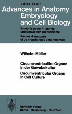
Studies on the structure and function of avian circumventricular organs in vitro (choroid plexus, glycogen body, pineal organ). The walls of the ventricular system in the brain show regional specializations termed circumventricular organs. Despite the fact that these organs have been sufficiently described morphologically, a convincing functional concept is lacking, especially in rela tion to adjacent nervous centers and to the cerebrospinal fluid. In vitro experimenta tion allows isolated analysis of circumventricular organs without interaction of other neuronal or humoral influences. Three avian circumventricular organs were selected for the present in vitro study: 1. choroid plexus, 2. glycogen body, and 3. pineal organ, all having the advantage of being anatomically well defined and therefore suited-for clean and easy isolation. Each of these represents an individual case with special prob-" lems for study in organ or cell culture. 1.1. Choroid Plexus In cultures of the choroid plexus the lack of vascular supply eliminates an important functional component which determines in vivo the polar organization of the plexus epithelium. Therefore, tissue culture studies are limited to processes occurring at the apical surface of the cell and focussed on the interaction between the cell and the culture medium. Choroid plexus from the lateral ventricles of 18 day-old chick em bryos were cultured successfully for up to 120 days in medium 199. In contrast, the choroid plexuses from the third and fourth ventricles required much greater amounts of media for effective cultivation.
Hinweis: Dieser Artikel kann nur an eine deutsche Lieferadresse ausgeliefert werden.
Hinweis: Dieser Artikel kann nur an eine deutsche Lieferadresse ausgeliefert werden.

