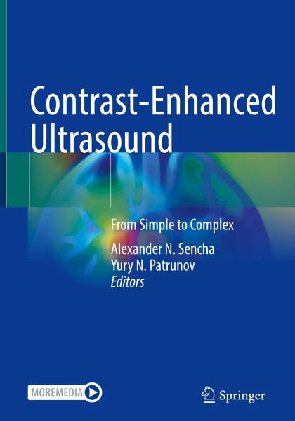
Contrast-Enhanced Ultrasound
From Simple to Complex
Herausgegeben: Sencha, Alexander N.; Patrunov, Yury N.

PAYBACK Punkte
57 °P sammeln!
This book provides a comprehensive analysis of the value of contrast-enhanced ultrasound (CEUS) in the diagnosis of a wide variety of pathologies. Sonography reliably identifies a wide range of diseases, and the efficacy of modern ultrasound has dramatically improved with contrast enhancement.This book covers almost all aspects of CEUS starting from basic principles and ending with features of its application in individual organs. In particular, it explores the diseases of abdominal, retroperitoneal, and pelvic organs as well as superficial structures, highlighting the characteristic features ...
This book provides a comprehensive analysis of the value of contrast-enhanced ultrasound (CEUS) in the diagnosis of a wide variety of pathologies. Sonography reliably identifies a wide range of diseases, and the efficacy of modern ultrasound has dramatically improved with contrast enhancement.
This book covers almost all aspects of CEUS starting from basic principles and ending with features of its application in individual organs. In particular, it explores the diseases of abdominal, retroperitoneal, and pelvic organs as well as superficial structures, highlighting the characteristic features of typical findings. Focal lesions are discussed in depth, with attention to their early detection and differential diagnosis. Besides, a practical approach to the stratification of the risk of malignancies is provided. The authors summarized their own experience with CEUS in oncology, hepatology, gynecology, urology, endocrinology, and other fields of medicine. The role ofCEUS in differential diagnosis of various disorders of the female reproductive system is comprehensively discussed as well. The presentation is clear and concise, and richly illustrated.
The book will be a helpful tool for both residents and practitioners approaching ultrasound diagnostics, as well for more experienced radiologists and other professionals.


