Conventional Radiography & CBCT in Localising Impacted Canines
Versandkostenfrei!
Versandfertig in 6-10 Tagen
36,99 €
inkl. MwSt.

PAYBACK Punkte
18 °P sammeln!
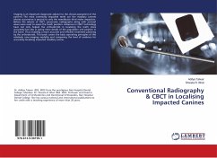
Imaging is an important diagnostic adjunct to the clinical assessment of the patient. The most commonly impacted teeth are the maxillary canines whose occurrence is second to only the mandibular third molar impaction. Before the advent of CBCT, panoramic radiographs along with occlusal views were used to assess the tooth position. Advances in CBCT technology have not only helped the orthodontist in localizing the teeth more accurately but also in giving more details of the angulation and position in the bone. Thus enabling a more accurate and efficient treatment planning by the orthodontist. T...
Imaging is an important diagnostic adjunct to the clinical assessment of the patient. The most commonly impacted teeth are the maxillary canines whose occurrence is second to only the mandibular third molar impaction. Before the advent of CBCT, panoramic radiographs along with occlusal views were used to assess the tooth position. Advances in CBCT technology have not only helped the orthodontist in localizing the teeth more accurately but also in giving more details of the angulation and position in the bone. Thus enabling a more accurate and efficient treatment planning by the orthodontist. This book covers the basic operating principles of this relatively new imaging modality and comparing the level of evidence for accurately localising impacted maxillary canine.


