Correction of Umbilical Hernias in Pigs with PVC
Umbilical Herniorrhaphy in Pigs
Versandkostenfrei!
Versandfertig in 6-10 Tagen
29,99 €
inkl. MwSt.

PAYBACK Punkte
15 °P sammeln!
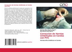
Two of the most common anatomical defects that occur in swine are scrotal hernias and umbilical hernias with a frequency of 1.7 to 6.7%. They are produced by weakening of the muscles around the umbilical stump, causing its opening with the intestines coming out and giving an appearance of mass in the anatomical area. The abdominal contents are enveloped by the parietal peritoneum. Diagnosis is basically clinical, with palpatory signs such as ring, continent and hernial contents, which are reduced with manual pressure. Radiology and ultrasonography are used as confirmatory techniques.Although t...
Two of the most common anatomical defects that occur in swine are scrotal hernias and umbilical hernias with a frequency of 1.7 to 6.7%. They are produced by weakening of the muscles around the umbilical stump, causing its opening with the intestines coming out and giving an appearance of mass in the anatomical area. The abdominal contents are enveloped by the parietal peritoneum. Diagnosis is basically clinical, with palpatory signs such as ring, continent and hernial contents, which are reduced with manual pressure. Radiology and ultrasonography are used as confirmatory techniques.Although there are several surgical techniques for correction, there is no one that is accepted as the best worldwide by surgeons. For this reason, new methods are always being sought that meet all expectations when performing herniorrhaphy, such as easy to practice, minimally invasive, effective, not requiring specialized instruments and demanding surgical environments, as well as biomaterials that meet some of the above requirements plus tissue acceptance at the site of implantation.


