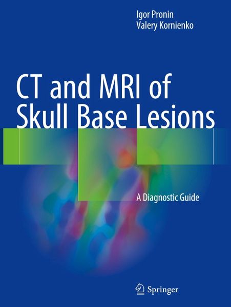
CT and MRI of Skull Base Lesions
A Diagnostic Guide

PAYBACK Punkte
61 °P sammeln!
This superbly illustrated book offers a comprehensive analysis of the diagnostic capabilities of CT and MRI in the skull base region with the aim of equipping readers with the knowledge required for accurate, timely diagnosis. The authors' vast experience in the diagnosis of skull base lesions means that they are ideally placed to realize this goal, with the book's contents being based on more than 10,000 histologically verified cases of frequent, uncommon, and rare diseases and disorders. In order to facilitate use, chapters are organized according to anatomic region. Readers will find clear ...
This superbly illustrated book offers a comprehensive analysis of the diagnostic capabilities of CT and MRI in the skull base region with the aim of equipping readers with the knowledge required for accurate, timely diagnosis. The authors' vast experience in the diagnosis of skull base lesions means that they are ideally placed to realize this goal, with the book's contents being based on more than 10,000 histologically verified cases of frequent, uncommon, and rare diseases and disorders. In order to facilitate use, chapters are organized according to anatomic region. Readers will find clear guidance on complex diagnostic issues and ample coverage of appearances on both standard CT and MRI methods and newer technologies, including especially CT perfusion, susceptibility- and diffusion-weighted MRI (SWI and DWI), and MR spectroscopy. The book will be an ideal reference manual for neuroradiologists, neurosurgeons, neurologists, neuro-ophthalmologists, neuro-otolaryngologists, craniofacial surgeons, general radiologists, medical students, and other specialists with an interest in the subject.


