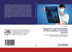
Diagnosis and localization of the maxillary impacted canines
By using multi-slice computed tomography and reconstructed panoramic radiograph
Versandkostenfrei!
Versandfertig in 6-10 Tagen
34,99 €
inkl. MwSt.

PAYBACK Punkte
17 °P sammeln!
The three dimensional CT images become superior to the conventional 2D radiographs for the localization of impacted canines and in the assessment of incisor root resorption by successessfully overcoming the limitations of the conventional radiography.This has the potential to allow for more thorough diagnosis, treatment planning, and assessment of changes over time and especially in the accurate localization of the impacted teeth including impacted canines, for that reason and because there is no previous Iraqi study on this subject the present study have been established to investigate with d...
The three dimensional CT images become superior to the conventional 2D radiographs for the localization of impacted canines and in the assessment of incisor root resorption by successessfully overcoming the limitations of the conventional radiography.This has the potential to allow for more thorough diagnosis, treatment planning, and assessment of changes over time and especially in the accurate localization of the impacted teeth including impacted canines, for that reason and because there is no previous Iraqi study on this subject the present study have been established to investigate with dental multi-slice computed tomography and 2D reconstructed panoramic like images, the locations of the impacted maxillary canines; the contact; overlapping; and resorption severity of the neighboring incisors. Furthermore this study was also performed to compare and evaluate whether there is any difference in the diagnostic information provided by the 3D multi-slice computed tomography (CT) and the 2D reconstructed panoramic like images in subjects with impacted maxillary canines.


