Michael W. Ross (Department of Clinical Studi Professor of Surgery, Sue J. Dyson (Centre for Equine Stud Head of Clinical Orthopaedics
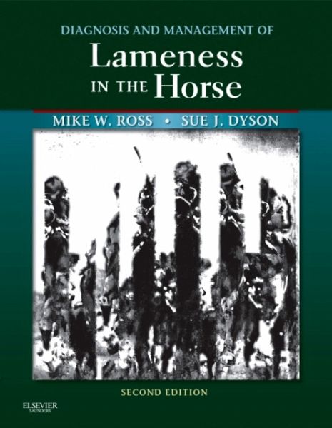
Diagnosis and Management of Lameness in the Horse
Michael W. Ross (Department of Clinical Studi Professor of Surgery, Sue J. Dyson (Centre for Equine Stud Head of Clinical Orthopaedics
Diagnosis and Management of Lameness in the Horse
- Gebundenes Buch
- Merkliste
- Auf die Merkliste
- Bewerten Bewerten
- Teilen
- Produkt teilen
- Produkterinnerung
- Produkterinnerung
Helping you to apply many different diagnostic tools, this book explores both traditional treatments and alternative therapies for conditions that can cause gait abnormalities in horses. It describes equine sporting activities and specific lameness conditions in major sport horse types.
Andere Kunden interessierten sich auch für
- Adams and Stashak's Lameness in Horses236,99 €
- Gary M. Baxter (Colorado State University, Fort Collins, Colorado)Manual of Equine Lameness99,99 €
- Juan C. Samper (Veterinary Reproductive Services, Langley, BC, CanaEquine Breeding Management and Artificial Insemination103,99 €
- Christopher C. Pollitt (Department of Co Reader in Equine MedicineThe Illustrated Horse's Foot139,99 €
- Equine Medicine, Surgery and Reproduction113,99 €
- Muir, William W., DVM, MSc, PhD, DACVA, DACVECC ( Regional DirectorEquine Anesthesia102,99 €
- C. Wayne McIlwraith (Barbara Cox University Distinguished ProfessorDiagnostic and Surgical Arthroscopy in the Horse214,99 €
-
-
-
Helping you to apply many different diagnostic tools, this book explores both traditional treatments and alternative therapies for conditions that can cause gait abnormalities in horses. It describes equine sporting activities and specific lameness conditions in major sport horse types.
Hinweis: Dieser Artikel kann nur an eine deutsche Lieferadresse ausgeliefert werden.
Produktdetails
- Produktdetails
- Verlag: Saunders
- Artikelnr. des Verlages: C2009-0-50774-X
- 2nd ed.
- Seitenzahl: 1424
- Erscheinungstermin: 13. Dezember 2010
- Englisch
- Abmessung: 284mm x 230mm x 53mm
- Gewicht: 3454g
- ISBN-13: 9781416060697
- ISBN-10: 1416060693
- Artikelnr.: 29957691
- Verlag: Saunders
- Artikelnr. des Verlages: C2009-0-50774-X
- 2nd ed.
- Seitenzahl: 1424
- Erscheinungstermin: 13. Dezember 2010
- Englisch
- Abmessung: 284mm x 230mm x 53mm
- Gewicht: 3454g
- ISBN-13: 9781416060697
- ISBN-10: 1416060693
- Artikelnr.: 29957691
1. Lameness Examination: Historical Perspective
2. Lameness in Horses: Basic Facts Before Starting
3. Anamnesis (History)
4. Conformation and Lameness
5. Observation: Symmetry and Posture
6. Palpation
7. Movement
8. Manipulation
9. Applied Anatomy of the Musculoskeletal System
10. Diagnostic Analgesia
11. Neurological Examination and Neurological Conditions Causing Gait Deficits
12. Unexplained Lameness
13. Assessment of Acute-Onset, Severe Lameness
14. The Swollen Limb
15. Radiography and Radiology
16. Ultrasonographic Evaluation of the Equine Limb: Technique
17. Ultrasonographic Examination of the Joints
18. Ultrasound and Orthopedic (Non-Articular) Disease
19. Nuclear Medicine
20. Computed Tomography
21. Magnetic Resonance Imaging
22. Gait Analysis for the Quantification of Lameness
23. Arthroscopic Examination
24. Tenoscopy and Bursoscopy
25. Themography: Use in Equine Lameness
26. Biomechanics of the Equine Limb and Its Effect on Lameness
27. The Foot and Shoeing
28. Trauma to the Sole and Wall
29. Functional Anatomy of the Palmar Aspect of the Foot
30. Navicular Disease
31. Fracture of the Navicular Bone and Congenital Bipartite Navicular Bone
32. Primary Lesions of the Deep Digital Flexor Tendon Within the Hoof Capsule
33. The Distal Phalanx and Distal Interphalangeal Joint
34. Laminitis
35. The Proximal and Middle Phalanges and Proximal Interphalangeal Joint
36. The Metacarpophalangeal Joint
37. The Metacarpal Region
38. The Carpus
39. The Antebrachium
40. The Elbow, Brachium, and Shoulder
41. The Hind Foot and Pastern
42. The Metatarsophalangeal Joint
43. The Metatarsal Region
44. The Tarsus
45. The Crus
46. The Stifle
47. The Thigh
48. Mechanical and Neurological Lameness in the Forelimbs and Hindlimbs
49. Diagnosis and Management of Pelvic Fractures in the Thoroughbred Racehorse
50. Lumbosacral and Pelvic Injuries in Sports and Pleasure Horses
51. Diagnosis and Management of Sacroiliac Joint Injuries
52. The Thoracolumbar Spine
53. The Cervical Spine and Soft Tissues of the Neck
54. Pathogenesis of Osteochondrosis
55. The Role of Nutrition in Developmental Orthopedic Disease: Nutritional Management
56. Diagnosis and Management of Osteochondrosis and Osseous Cyst-like Lesions
57. Physitis
58. Angular Limb Deformitis
59. Flexural Limb Deformity in Foals
60. Cervical Stenotic Myelopathy
61. Osteoarthritis
62. Markers of Osteoarthritis: Implications for Early Diagnosis and Monitoring of Pathology and Effects of Therapy
63. Gene Therapy
64. Models of Equine Joint Disease
65. Infectious Arthritis
66. Non-infectious Arthritis
67. Joint Conditions
68. Pathophysiology of Tendon Injury
69. Superficial Digital Flexor Tendonitis
70. The Deep Digital Flexor Tendon
71. Desmitis of the Accessory Ligament of the Deep Digital Flexor Tendon
72. The Suspensory Apparatus
73. Clinical Use of Stem Cells, Marrow Components, and Other Growth Factors
74. Diseases of the Digital Synovial Sheath, Palmar Annular Ligament, and Digital Annular Ligaments
75. The Carpal Canal and The Carpal Synovial Sheath
76. The Tarsal Sheath
77. Extensor Tendon Injury
78. Curb
79. Bursae and Other Soft Tissue Swellings
80. Other Soft Tissue Injuries
81. Tendon Lacerations
82. Soft Tissue Injuries of the Pastern
83. Skeletal Muscle and Lameness
84. Principles and Practice of Joint Disease Treatment
85. Analgesia and Hindlimb Lameness
86. Bandaging, S