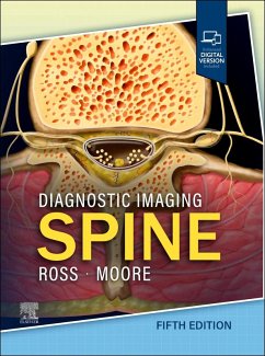
Covering the entire spectrum of this fast-changing field, Diagnostic Imaging: Spine, fifth edition, is an invaluable resource for general radiologists, neuroradiologists, and trainees-anyone who requires an easily accessible, highly visual reference on today’s spinal imaging. Drs. Jeffrey Ross, Kevin Moore, and their team of highly regarded experts provide updated information on disease identification and imaging techniques to help you make informed decisions at the point of care. The text is lavishly illustrated, delineated, and referenced, making it a useful learning tool as well as a handy reference for daily practice. * Serves as a one-stop resource for key concepts and information on radiologic imaging and interpretation of the spine, neck, and central nervous system * Contains six robust sections, each beginning with normal imaging anatomy and covering all aspects of this challenging field: Congenital and Genetic Disorders, Trauma, Degenerative Diseases and Arthritides, Infection and Inflammatory Disorders, Peripheral Nerve and Plexus, and Spine Postprocedural/Posttreatment Imaging * Features 3,200 high-quality print images (with an additional 2,000 images in the complimentary eBook), including radiologic images, full-color medical illustrations, clinical photographs, histologic images, and gross pathologic photographs * Provides new and expanded content on CSF leak disorder and root sleeve leak; CSF-venous fistulas; demyelinating disease based upon better knowledge of MS; neuromyelitis optica spectrum disorder; anti-MOG disorders; malignant nerve sheath tumor and paragangliomas; and spinal ependymomas, including myxopapillary and classical cellular spinal ependymoma * Contains new chapters on both imaging technique and diseases/disorders, and existing chapters have been rearranged to better represent current information on inflammatory and autoimmune disorders and systemic manifestations of diseases * Provides updates from cover to cover, including overviews and new recommendations for evaluation of transitional spinal anatomy (spine enumeration), which have important and practical applications in routine imaging with downstream effects on spine intervention * Uses bulleted, succinct text and highly templated chapters for quick comprehension of essential information at the point of care * Includes an eBook that allows you access to everything in the print version as well as additional images, text, and references, with the ability to search, customize your content, make notes and highlights, and have content read aloud; additional digital ancillary content may publish up to 6 weeks following the publication date ¿ Overview, update, and recommendations for evaluation of transitional spinal anatomy (spine enumeration). This has important and practical application in routine imaging with downstream effects on spine intervention. Specific recommendations are given ¿ Update on treatments for CSF-venous fistulas including venous embolization and percutaneous fibrin glue placement. Updates on new technology, such as photon-counting CT that have direct application for diagnosis of this pathology. ¿ Updates on spinal tumor WHO classification, specifically spinal ependymomas including myxopapillary and classical cellular spinal ependymoma ¿ Updates on classification of myelofibrosis and myelodysplastic syndromes ¿ Updates on classification of demyelinating disorders including MOGAD and NMOSD ¿ Update and expansion for classification of congenital spinal open and closed neural tube defects
Hinweis: Dieser Artikel kann nur an eine deutsche Lieferadresse ausgeliefert werden.
Hinweis: Dieser Artikel kann nur an eine deutsche Lieferadresse ausgeliefert werden.


