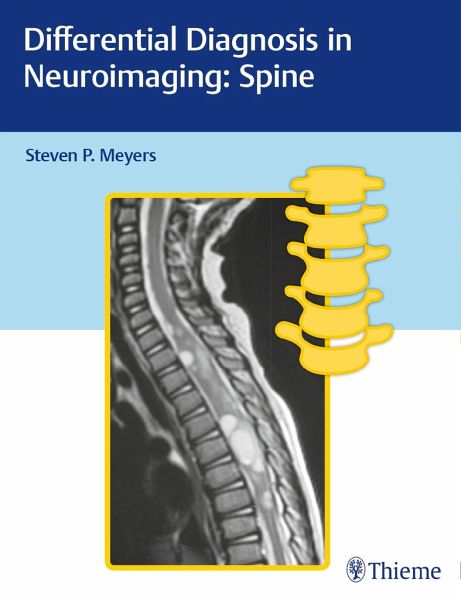
Differential Diagnosis in Neuroimaging: Spine

PAYBACK Punkte
53 °P sammeln!
A unique, easy-to-use, and definitive guide to diagnostic spine imagingAuthored by renowned neuro-radiologist Steven P. Meyers, Differential Diagnosis in Neuroimaging: Spine is a stellar guide for identifying and diagnosing cervical, thoracic, lumbar, and sacral spine anomalies based on location and neuroimaging results. The succinct text reflects more than 25 years of hands-on experience gleaned from advanced training and educating residents and fellows in radiology, neurosurgery, and orthopaedic surgery. The high-quality MRI, CT, and X-ray images have been collected over Dr. Meyers's lengthy...
A unique, easy-to-use, and definitive guide to diagnostic spine imaging
Authored by renowned neuro-radiologist Steven P. Meyers, Differential Diagnosis in Neuroimaging: Spine is a stellar guide for identifying and diagnosing cervical, thoracic, lumbar, and sacral spine anomalies based on location and neuroimaging results. The succinct text reflects more than 25 years of hands-on experience gleaned from advanced training and educating residents and fellows in radiology, neurosurgery, and orthopaedic surgery. The high-quality MRI, CT, and X-ray images have been collected over Dr. Meyers's lengthy career, presenting an unsurpassed visual learning tool.
The distinctive 'three-column table plus images' format is easy to incorporate into clinical practice, setting this book apart from larger, disease-oriented radiologic tomes. This layout enables readers to quickly recognize and compare abnormalities based on high-resolution images.
Key Highlights
- Tabular columns organized by anatomical abnormality include imaging findings and a summary of key clinical data that correlates to the images
- Congenital/developmental abnormalities, spinal deformities, and acquired pathologies in both children and adults
- Lesions organized by region including dural, intradural extramedullary, extra-dural, and sacrum
- More than 600 figures illustrate the radiological appearance of spinal tumors, lesions, deformities, and injuries
- Spinal cord imaging for the diagnosis of intradural intramedullary lesions and spinal trauma
This visually rich resource is a must-have diagnostic tool for trainee and practicing radiologists, neurosurgeons, neurologists, physiatrists, and orthopaedic surgeons who specialize in treating spine-related conditions. The highly practical format makes it ideal for daily rounds, as well as a robust study guide for physicians preparing for board exams.


