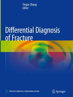
This book covers diagnostic images of common and rare fractures for nearly every part of the human body, based on a large number of clinical cases. The highlight of this book is that both of three-dimensional X-ray images and CT/MRI images of thousands of fracture cases are presented for comparison and further discussion, according to the framework of AO classification.
The first chapter gives a general introduction of various diagnostic imaging techniques for fractures, with attention to their advantages and disadvantages. The following chapters present detailed radiological images of upper extremity fractures, lower extremity fractures, axial skeleton fractures, and epiphyseal lesions. It helps readers to recognize the difference between various diagnostic techniques, and to select optimal imaging techniques. With the illustrative figures, this book is a valuable tool to orthopaedist, radiologists, trauma surgeons, emergency room doctors, professional clinical staff, and medical students.
Hinweis: Dieser Artikel kann nur an eine deutsche Lieferadresse ausgeliefert werden.
"The images and illustrations used throughout are excellent and clear, with well labelled detail associated with each image. There is a wide selection of imaging used in the book, such as radiography (x-ray), CT and MR ... . The lasting impression of this textbook on the reader is the excellent, detailed and clearly labelled images with supporting anatomical infographics." (Lisa Field, RAD Magazine, September, 2022)

