Digital Imaging in Conservative Dentistry and Endodontics
Versandkostenfrei!
Versandfertig in 6-10 Tagen
41,99 €
inkl. MwSt.

PAYBACK Punkte
21 °P sammeln!
Radiographs are the most accurate and least subjective diagnostic aids available to endodontists for diagnosis of diseases affecting maxilla and mandible. Digital imaging incorporates computer technology in the capture, display, enhancement and storage of direct radiographic images. Unfortunately, the early systems could not capture panoramic and cephalometric images, and this made it impossible for surgeries to abandon film processing and adopt digital technology. Digitalization of dental records, computer assisted imaging techniques and virtual treatment planning or simulations, have revolut...
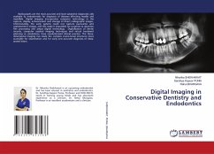
Radiographs are the most accurate and least subjective diagnostic aids available to endodontists for diagnosis of diseases affecting maxilla and mandible. Digital imaging incorporates computer technology in the capture, display, enhancement and storage of direct radiographic images. Unfortunately, the early systems could not capture panoramic and cephalometric images, and this made it impossible for surgeries to abandon film processing and adopt digital technology. Digitalization of dental records, computer assisted imaging techniques and virtual treatment planning or simulations, have revolutionized clinical practice. The three- dimensional imaging has made the complex cranio-facial structures more accessible for examination and for early and accurate diagnosis of deep-seated lesion.


