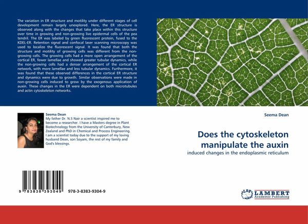
Does the cytoskeleton manipulate the auxin
induced changes in the endoplasmic reticulum
Versandkostenfrei!
Versandfertig in 6-10 Tagen
32,99 €
inkl. MwSt.

PAYBACK Punkte
16 °P sammeln!
The variation in ER structure and motility under different stages of cell development remain largely unexplored. Here, the ER structure is observed along with the changes that take place within this structure over time in growing and non-growing live epidermal cells of the pea tendril. The ER was labeled by green fluorescent protein, fused to the KDEL-ER. Retention signal and confocal laser scanning microscopy was used to localize the fluorescent signal. It was found that both the structure and motility of growing cells was different from the non-growing cells. The growing cells had a more ope...
The variation in ER structure and motility under different stages of cell development remain largely unexplored. Here, the ER structure is observed along with the changes that take place within this structure over time in growing and non-growing live epidermal cells of the pea tendril. The ER was labeled by green fluorescent protein, fused to the KDEL-ER. Retention signal and confocal laser scanning microscopy was used to localize the fluorescent signal. It was found that both the structure and motility of growing cells was different from the non-growing cells. The growing cells had a more open arrangement of the cortical ER, fewer lamellae and showed greater tubular dynamics, while the non-growing cells had a denser arrangement of the cortical ER network, with more lamellae and less tubular dynamics. Furthermore, it was found that these observed differences in the cortical ER structure and dynamics were due to growth. Similar observations were made in non-growing cells induced to grow by the exogenous application of auxin. These changes in the ER were dependent on both microtubules and actin cytoskeleton networks.


