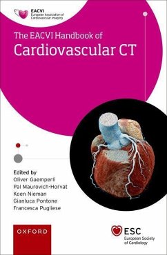
Eacvi Handbook of Cardiovascular CT
Herausgeber: Gaemperli, Oliver; Pugliese, Francesca; Pontone, Gianluca; Nieman, Koen; Maurovich- Horvat, Pál
Eacvi Handbook of Cardiovascular CT
Herausgeber: Gaemperli, Oliver; Pugliese, Francesca; Pontone, Gianluca; Nieman, Koen; Maurovich- Horvat, Pál
- Broschiertes Buch
- Merkliste
- Auf die Merkliste
- Bewerten Bewerten
- Teilen
- Produkt teilen
- Produkterinnerung
- Produkterinnerung
The handbook represents an important step towards dissemination of skills and knowledge in cardiovascular CT. It is a concise and practical companion, to benefit students, trainees or advanced users; cardiologists, radiologists, cardiac surgeons or technicians, in their everyday practice.
Andere Kunden interessierten sich auch für
![The Eacvi Handbook of Nuclear Cardiology The Eacvi Handbook of Nuclear Cardiology]() Alessia GimelliThe Eacvi Handbook of Nuclear Cardiology52,99 €
Alessia GimelliThe Eacvi Handbook of Nuclear Cardiology52,99 €![The Eacvi Echo Handbook The Eacvi Echo Handbook]() The Eacvi Echo Handbook61,99 €
The Eacvi Echo Handbook61,99 €![Orbit and Neuro-Ophthalmic Imaging, an Issue of Neuroimaging Clinics Orbit and Neuro-Ophthalmic Imaging, an Issue of Neuroimaging Clinics]() Juan E. GutierrezOrbit and Neuro-Ophthalmic Imaging, an Issue of Neuroimaging Clinics114,99 €
Juan E. GutierrezOrbit and Neuro-Ophthalmic Imaging, an Issue of Neuroimaging Clinics114,99 €![Esc Textbook of Cardiovascular Nursing Esc Textbook of Cardiovascular Nursing]() Esc Textbook of Cardiovascular Nursing80,99 €
Esc Textbook of Cardiovascular Nursing80,99 €![Pet/CT in Cancer: An Interdisciplinary Approach to Individualized Imaging Pet/CT in Cancer: An Interdisciplinary Approach to Individualized Imaging]() Mohsen BeheshtiPet/CT in Cancer: An Interdisciplinary Approach to Individualized Imaging116,99 €
Mohsen BeheshtiPet/CT in Cancer: An Interdisciplinary Approach to Individualized Imaging116,99 €![Introduction to Intra-Operative and Surgical Radiography Introduction to Intra-Operative and Surgical Radiography]() Jim Hughes (CT Radiographer, CT Radiographer, Leeds Teaching HospitIntroduction to Intra-Operative and Surgical Radiography64,99 €
Jim Hughes (CT Radiographer, CT Radiographer, Leeds Teaching HospitIntroduction to Intra-Operative and Surgical Radiography64,99 €![Cardiac Catheterization and Coronary Intervention Cardiac Catheterization and Coronary Intervention]() Cardiac Catheterization and Coronary Intervention74,99 €
Cardiac Catheterization and Coronary Intervention74,99 €-
-
-
The handbook represents an important step towards dissemination of skills and knowledge in cardiovascular CT. It is a concise and practical companion, to benefit students, trainees or advanced users; cardiologists, radiologists, cardiac surgeons or technicians, in their everyday practice.
Hinweis: Dieser Artikel kann nur an eine deutsche Lieferadresse ausgeliefert werden.
Hinweis: Dieser Artikel kann nur an eine deutsche Lieferadresse ausgeliefert werden.
Produktdetails
- Produktdetails
- The European Society of Cardiology Series
- Verlag: Oxford University Press
- Seitenzahl: 384
- Erscheinungstermin: 18. Februar 2023
- Englisch
- Abmessung: 195mm x 129mm x 18mm
- Gewicht: 410g
- ISBN-13: 9780192884459
- ISBN-10: 019288445X
- Artikelnr.: 66408715
- Herstellerkennzeichnung
- Libri GmbH
- Europaallee 1
- 36244 Bad Hersfeld
- gpsr@libri.de
Prof Gaemperli studied medicine at the University of Zurich and graduated in 2003. From 2003 to 2009 he underwent a clinical fellowship in internal medicine and cardiology at the University Hospital Zurich. From 2009 to 2010, Prof. Gaemperli joined the MRC Cyclotron Unit of the Hammersmith Hospital in London, UK for a 2-year research fellowship in cardiac PET, MRI and CT-Imaging. After this, he returned to the University Hospital Zurich where he assummed a Consultant Position and was trained in Interventional Cardiology. In 2016, he was awarded with the Sheikh Khalifa Professorship for Interventional Cardiology and Cardiac Imaging. His main fields of interest are Cardiac Imaging and interventional cardiology. Since 2018, Prof. Gaemperli holds a position as consultant interventional cardiologist and CEO of the HeartClinic Hirslanden Zurich. Pál Maurovich-Horvat MD PhD MPH, Associate Professor of Cardiology and Radiology, director of the Cardiovascular Imaging Research Group at the Semmelweis University in Budapest, Hungary. He is the president of the Hungarian Association of Cardiovascular Imaging, and board member of the European Association of Cardiovascular Imaging of the European Society of Cardiology Dr. Maurovich-Horvat graduated from the Semmelweis University in 2006, which was was followed by a three-year long advanced cardiovascular imaging research fellowship at Massachusetts General Hospital and Harvard Medical School. Dr. Maurovich-Horvat studied clinical effectiveness at the Harvard University, School of Public Health, where he graduated in 2012. His research interest focuses on the development of non-invasive diagnostic imaging strategies to identify high-risk patients and vulnerable coronary plaques. Dr Nieman is a cardiologist and professor in the departments of medicine/cardiovascular and radiology. He investigates advanced cardiac imaging techniques. He is currently the president of the Society of Cardiovascular Computed Tomography. Dr Nieman was born in the Netherlands, obtained his medical degree at the Radboud University in Nijmegen (1998), and completed his cardiology training at the Erasmus University Medical Center in Rotterdam (2008). His research in cardiac CT at the Erasmus University resulted in a PhD degree in 2003. In 2004 he performed an imaging fellowship at the Massachusetts General Hospital (Harvard Medical School) in Boston, MA. Dr Nieman joined the staff of the department of cardiology and radiology at the Erasmus University Medical Center in 2008, where he was scientific director of the cardiac CT and MRI group and supervised the intensive cardiac care unit until he joined the staff at Stanford University. Dr Gianluca Pontone was graduated with honors in medicine in 1997 followed by post-graduate degree in cardiology and radiology and PhD in Clinical Methodology in 2001, 2006 and 2014 at University of Milan, Italy. He is currently director of Cardiovascular Imaging Department of Centro Cardiologico Monzino, a fully dedicated research hospital to cardiovascular disease where the University of Milan is based. He is author of more than 300 indexed articles on international journal, several scientific books, more than 500 scientific abstracts and lectures at national and international meetings in the field of cardiovascular imaging. He is currently in the board member of SCCT, EACVI, chairman of CT certification committee of European Association of Cardiovascular Imaging (EACVI), past chairman of working group of cardiac magnetic resonance of Italian society of cardiology and deputy chairman of working group of cardiac computed tomography of Italian society of Cardiology. Francesca Pugliese qualified from Medical School of University of Genoa in 2000 and was awarded her certificate of completion of speciality training in 2004. She then joined the non-invasive cardiac imaging group at the Thoraxcentre/Erasmus MC University Medical Centre Rotterdam and received her PhD with honors in 2008. Since then she has worked for the Royal Brompton hospital and in the Cardiology group of the Medical Research Council (MRC) Clinical Sciences Centre, Imperial College London, at Hammersmith Hospital, in cardiovascular hybrid imaging (PET/CT). After contributing to the clinical service of the Essex Cardiothoracic Centre in Essex in 2009, she joined the Centre for Advanced Cardiovascular Imaging and the NIHR Cardiovascular Biomedical Research Unit at Barts as a Senior Clinical Lecturer in May 2010.
* 1.Technical background, patient preparation, quality/safety
* 1.1: Marcel van Straten, Sebastian Vandermolen, and Francesca
Pugliese: Key hardware components
* 1.2: Ulrike Haberland and Thomas Allmendinger: Technical
specifications
* 1.3: Alexia Rossi, Martina DeKnegt, and Jens Hove: Physical
background of radiation
* 1.4: Ronak Rajani and Mihaly Karolyi: Patient selection and
preparation
* 1.5: Ulrike Haberland, Thomas Allmendinger, and Francesca Pugliese:
Scanner setup and image acquisition protocols
* 1.6: Jamal Khan, Sarah Moharem-Elgamal, and Francesca Pugliese: Image
reconstruction, post-processing and analysis
* 1.7: Anna Beattie and Francesca Pugliese: Radiation exposure
* 1.8: Oliver Gaemperli: Image artifacts
* 1.9: Mohamed Marwan and Mihaly Karolyi: Tips and tricks to improve
image quality
* 1.10: Casper Mihl and Bibi Martens: Contrast agents and injection
protocols
* 1.11: Ricardo Budde, Sarah Moharem-Elgamal, and Francesca Pugliese:
Challenging scenarios
* 2. Indications: Coronary artery disease
* 2.1: Martin Willemink: Coronary artery calcium Imaging
* 2.2: Balint Szilveszter, Csilla Celeng, Richard Takx, and Pál
Maurovich-Horvat: Coronary CT angiography interpretation and
reporting
* 2.3: Tessa Genders: Patients with chronic coronary syndrome
* 2.4: Domenico Mastrodicasa: CT-based fractional flow reserve
* 2.5: Koen Nieman and Gianluca Pontone: Myocardial perfusion and scar
imaging
* 2.6: Marton Kolossvary: Atherosclerotic plaque imaging
* 2.7: Sujana Balla and Koen Nieman: Stents and bypass grafts
* 2.8: Andrew Chang, Ian Rogers, and Koen Nieman: Coronary anomalies
* 2.9: Admir Dedic and Murat Arslan: Patients with acute coronary
syndrome
* 2.10: Andrea Bartykowszki: Graft vasculopathy in transplanted hearts
* 3. Non-CAD indications
* 3.1: Richard Takx and Csilla Celeng: Ventricular dimensions and
function
* 3.2: Giuseppe Muscogiuri: Cardiac reference values
* 3.3: Giuseppe Muscogiuri: Aortic valve disease
* 3.4: Marco Guglielmo: Preprocedural aortic valve assessment
* 3.5: Marco Guglielmo: Mitral valve disease
* 3.6: Andrea Baggiano: Postprocedural valve assessment
* 3.7: Marco Guglielmo: Left atrium and pulmonary veins
* 3.8: Andrea Baggiano: Infective endocarditis
* 3.9: Michael Messerli: Great thoracic vessels
* 3.10: Andreas Giannopoulos: Adult congenital heart disease
* 3.11: Dominik Benz: Cardiomyopathies
* 3.12: Andreas Giannopoulos: Cardiac masses
* 3.13: Sarah Moharem: Pericardial disease
* 3.14: Dominik Benz: Implanted cardiac devices
* 3.15: Michael Messerli: Extracardiac findings
* 3.16: Marton Kolossvary: Artificial intelligence
* 4. Training and competence in cardiac CT
* 4.1: Andrea Baggiano: EACVI certification standards in Cardiac CT

