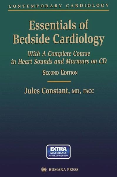
Essentials of Bedside Cardiology, Second Edition, like the first edition, is designed for those who wish to balance technological advances with increased personal skill in history taking and physical examination. It is important to teach physicians that all technologies now in use for diagnosing cardiovascular disorders, such as echocardiography, can have false positive and false negative results. It is not always wise to rely on these technologies alone; indeed, they may not even be available in some settings. Even when the full panoply of up-to-date techniques is at the physician's disposal,…mehr
Essentials of Bedside Cardiology, Second Edition, like the first edition, is designed for those who wish to balance technological advances with increased personal skill in history taking and physical examination. It is important to teach physicians that all technologies now in use for diagnosing cardiovascular disorders, such as echocardiography, can have false positive and false negative results. It is not always wise to rely on these technologies alone; indeed, they may not even be available in some settings. Even when the full panoply of up-to-date techniques is at the physician's disposal, the patient may not be a good candidate for an echocardiogram, or the technician or reader may not be well qualified, or the equipment itself may be substandard. Technology must be combined with physical examination to decide what is true and what is false. The practice of expert history taking and physical examination returns the physician to the actual patient, where the physician can feel likea "real doctor" rather than a mere interpreter of laboratory data. Essentials of Bedside Cardiology, Second Edition, strives to teach and not simply to tell the facts, relying on three basic methods derived from the psychology of teaching and learning: 1. Explain the facts. 2. Use a question and answer format-the Socratic method. 3. Provide tricks or mnemonics to help the reader remember the facts.
1 The Checklist in History Taking.- Advantages of a Checklist.- Reminder List History.- Etiologies.- Follow-Up Questions.- New York Heart Association Functional and Therapeutic Classification.- 2 Cardiac Clues from Physical Appearance.- Skin.- Congenital and Acquired Facies.- Eyes.- Edema.- Extremities.- Chest and Respiration.- 3 Arterial Pulses and Pressures.- Method of Arm Palpation.- Rates of Rise and Pulse Volume.- Pulsus Bisferiens.- Palpation of the Leg Pulses.- Estimating Systolic Blood Pressure by Palpation Alone.- Accurate Blood Pressure Measurement.- Korotkoff Sounds.- Chest Piece Placement and Choice.- Equipment Sources of Error.- Systolic Pressure Measurement.- Diastolic Blood Pressure Recording.- Accuracy of Blood Pressure Recording.- Summary of How to Take Arm Blood Pressures by Listening for Korotkoff Sounds.- Pseudohypertension.- Pulsus Alternans.- Blood Pressure and Pulses in the Legs.- Pulsus Paradoxus.- Capillary Pulsation.- Abdominal Aortic Aneurysm (AAA).- How to Tell Cardiac Function by Blood Pressure Response to a Valsalva Maneuver.- 4 Jugular Pressure and Pulsations.- Venous Pressure by Jugular Inspection.- How to Use the Internal Jugulars as a Manometer.- Method of Obtaining an Accurate Measurement of Venous Pressure.- How to Tell Jugular from Carotid Pulsations.- The Abdominal Compression Test (Hepatojugular Reflux).- Jugular Pulse Contours.- 5 Inspection and Palpation of the Chest.- Ventricular Enlargement from Examination of the Chest.- Cardiac Dilatation Signs in the Left Lateral Decubitis Position.- Right Ventricular Enlargement.- Left-Sided Causes of Left Parasternal Movement.- X-ray Evaluation of Cardiac Size.- Diagnosis of Ventricular Hypertrophy by Physical Examination of the Precordium.- 6 The Stethoscope.- The Bell Chest Piece.- The Smooth Diaphragm.- The Tubing.- Air Leaks and Ear Tips.- Summary of Good Stethoscope Characteristics.- 7 Diagramming and Grading Heart Sounds and Murmurs.- The Auscultogram.- Grading of Heart Sounds and Murmurs.- 8 The First Heart Sound (S1).- Physiology of the First Sound Components.- The M1 Plus Aortic Ejection Sound as the Cause of a Split S1.- The Pulmonary Ejection Sound.- Loudness of the M1.- 9 The Second Heart Sound (S2).- Physiology and Nomenclature for the S2.- Explanation of Normal Splitting Sequence of S2.- Physiology of the Normally Moving Split.- Loudness of the Components of S2.- The Widely Split S2.- The A2-P2 in Pulmonary Stenosis.- ASDs with Narrow Splits.- Differential Diagnosis of the Fixed Split.- The Narrowly Split S2.- The S2 Split in Pulmonary Hypertension.- The Reversed or Paradoxically Split S2.- 10 The Opening Snap.- Mechanism and Timing.- Relation Between the 2-OS Interval and the Severity of Mitral Stenosis.- The Loudness of the Opening Snap.- How to Tell an A2-P2 from an A2-OS.- 11 The Third Heart Sound (S3).- Nomenclature.- Timing.- Mechanism of Production.- The Physiological S3.- Loudness of the S3.- The Exaggerated Physiological S3.- The PathologicalS3.- The Physiological Versus the Pathological S3.- The Right Versus the Left Ventricular S3.- The S3 Versus the Opening Snap.- 12 The Fourth Heart Sound (S4).- Nomenclature.- Mode of Production.- Recognizing the Rhythm of the S4 Gallop.- The Physiological S4.- The Pathological S4.- Loudness and Audibility of the S4.- Differentiation of the S4 from the S3.- Differentiation of an S4-S1 from a Split S1 (M1-A1 or M1-T1).- Severity of Cardiac Dysfunction and Presence of an S4.- The S4-S1 Interval and Severity of Dysfunction.- Summation and Augmented Gallops.- The Pacemaker S4-Like Click.- 13 Ejection Murmurs.- Physical Causes.- Characteristics of the Ejection Murmur.- Types of Ejection Murmurs.- Systolic Flow Murmurs.- Aortic Stenosis Ejection Murmurs.- Pulmonary Stenosis Ejection Murmurs.- The Effect of Respiration on PS Ejection Murmurs.- Thoracic, Subclavian, Carotid, and Thyroid Artery Flow Murmurs.- 14 Systolic Regurgitant Murmurs.- The Effect of a Sudden Long Diastole on Left-Sided Regurgitant Murmurs.- Mitral Regurgitation Murmurs.- Effect of Drugs and Maneuvers on Systolic Regurgitant Murmurs.- The Prolapsed Mitral Valve Syndrome (Barlow's Syndrome).- Tricuspid Regurgitation (TR) Murmurs.- The Cardiorespiratory Murmur.- VSD Murmurs.- Continuous Murmurs.- 15 Diastolic Murmurs.- Diastolic Atrioventricular Valve Murmurs.- Tricuspid Diastolic Flow Murmurs.- Tricuspid Stenosis (TS) Diastolic Murmurs.- Diastolic Semilunar Valve Murmurs.- Pulmonary Regurgitation Murmurs.- Pericardial Friction Rubs.- 16 Abdominal Murmurs.- Normal Abdominal Murmurs.- Abnormal Abdominal Murmurs.
Rezensionen
"This is a thoughtful book, particularly strong on the correct physiological explanation of physical signs. I have always found Jules Constant's book to be my first (and only) reference regarding physical signs, and it is a great pleasure to see this updated version. Given the breadth of coverage it is surprisingly short and readable, and highly recommended." -Cardiology News
Es gelten unsere Allgemeinen Geschäftsbedingungen: www.buecher.de/agb
Impressum
www.buecher.de ist ein Shop der buecher.de GmbH & Co. KG i.I. Bürgermeister-Wegele-Str. 12, 86167 Augsburg Amtsgericht Augsburg HRA 13309