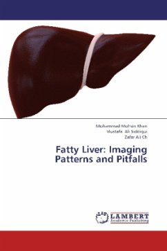
Fat accumulation is one of the most common abnormalities of the liver depicted on cross-sectional images. Common patterns include diffuse fat accumulation, diffuse fat accumulation with focal sparing, and focal fat accumulation in an otherwise normal liver. Unusual patterns that may cause diagnostic confusion by mimicking neoplastic, inflammatory, or vascular conditions include multinodular and perivascular accumulation. All of these patterns involve the heterogeneous or nonuniform distribution of fat. To help prevent diagnostic errors and guide appropriate work-up and management, radiologists should be aware of the different patterns of fat accumulation in the liver, especially as they are depicted at ultrasonography, computed tomography, and magnetic resonance imaging. In addition, knowledge of the risk factors and the pathophysiologic, histologic, and epidemiologic features of fat accumulation may be useful for avoiding diagnostic pitfalls and planning an appropriate work-up in difficult cases.
Bitte wählen Sie Ihr Anliegen aus.
Rechnungen
Retourenschein anfordern
Bestellstatus
Storno

