Fundamentals of Medical Imaging Systems
Versandkostenfrei!
Versandfertig in über 4 Wochen
136,99 €
inkl. MwSt.

PAYBACK Punkte
68 °P sammeln!
The method and practice of functional and structural imaging of the interior of a body for the purpose of clinical examination and subsequent medical intervention is referred to as medical imaging. Medical imaging techniques help in creating visual representation of the structure and function of different body parts, organs or tissues. They have proved to be extremely beneficial in diagnosing and curing diseases. They also help in creating a normal anatomy and physiology database, allowing the detection of abnormalities. In medical imaging, two types of radiographic images are used: projection...
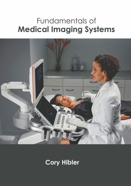
The method and practice of functional and structural imaging of the interior of a body for the purpose of clinical examination and subsequent medical intervention is referred to as medical imaging. Medical imaging techniques help in creating visual representation of the structure and function of different body parts, organs or tissues. They have proved to be extremely beneficial in diagnosing and curing diseases. They also help in creating a normal anatomy and physiology database, allowing the detection of abnormalities. In medical imaging, two types of radiographic images are used: projection radiography and fluoroscopy. Fluoroscopy, like radiography, produces real-time images of internal body structures, but it uses a steady input of X-rays at a lower dose rate. X-rays, or projectional radiographs, are often used to diagnose the kind and extent of a fracture as well as to detect pathological changes in the lungs. X-rays can be used to visualize the structure of the stomach and intestines, which is helpful in diagnosing ulcers and some types of colon cancer. This book consists of contributions made by international experts. Also included herein is a detailed explanation of the various concepts and applications of medical imaging systems. For someone with an interest and eye for detailed research, this book covers the most significant topics in this field.


