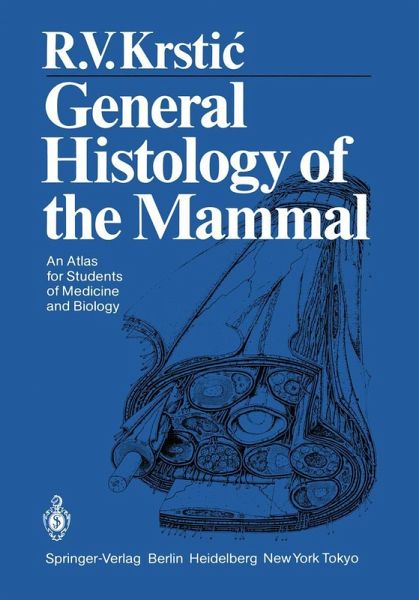
In this book, concerned with the spatial and structural repre sentation of the cell and its particular specializations, the author hasdeviatedconsiderablyfrom the usual planfor other books related to this subject and has presented the illustrative material in the form of detailed and accurate drawings. The layout of the book provides the reader with a briefnarrative accountoftheparticularorganelleaccompanied byafull-plate illustration on the facing page. Most ofthe narrative accounts areaccompaniedbyashortbibliographyofgermanereferences in the event the reader desires to pursue the subject…mehr
In this book, concerned with the spatial and structural repre sentation of the cell and its particular specializations, the author hasdeviatedconsiderablyfrom the usual planfor other books related to this subject and has presented the illustrative material in the form of detailed and accurate drawings. The layout of the book provides the reader with a briefnarrative accountoftheparticularorganelleaccompanied byafull-plate illustration on the facing page. Most ofthe narrative accounts areaccompaniedbyashortbibliographyofgermanereferences in the event the reader desires to pursue the subject matter in greater depth. In my estimation there is no other presentation currentlyavailablewhich utilizes thisapproach to demonstrate the cellular components and their associated morphophysi ology with such elegance. Thetextisclearlywrittenand,althoughtheindividualaccounts are brief, they are highly informative with all ofthe important details being provided. The accuracy ofthe textural presenta tions and the continuity ofexpression provide strong evidence that the author has spent an enormous amount of time in preparing the text and in painstakingly drawing the illustra tions. Certainly, an obvious strength ofthis volume is the high qualityoftheillustrativerenditions, allofwhichweredrawnby Dr. KRSTIC. These attest to the profound and comprehensive nature of the author's knowledge of the field of cellular and structural biology. This book is truly a work oflove and art by one who is gifted both didactically and artistically. Dr. KRSTIC should be con gratulated for providing us with an extremely accurate and detailedaccountofthe 2-dimensional, andinmanycasesthe 3 dimensional, view of the cell and its component organelles. It has no equal.Hinweis: Dieser Artikel kann nur an eine deutsche Lieferadresse ausgeliefert werden.
I. Introduction.- Origin of Tissues.- Differentiation. Closed and Wide-Meshed Cell Unions.- Four Tissue Groups.- Growth, Regeneration, Hyperplasia, Hypertrophy, Atrophy, Involution, Degeneration, and Necrosis.- Metaplasia.- Transplantation, Implantation, and Explantation.- II. Epithelial Tissue.- Surface Epithelia Classification of Surface Epithelia.- Localization of Various Surface Epithelia.- Simple Squamous Epithelium.- Simple Cuboidal Epithelium.- Simple Columnar Epithelium.- Pseudostratified Columnar Epithelium.- Transitional Epithelium.- Stratified Columnar Epithelium.- Nonkeratinized Stratified Squamous Epithelium.- Keratinized Stratified Squamous Epithelium (Epidermis).- Secretory Surface Epithelia Amniotic Epithelium.- Vascularized Secretory Epithelium.- Atypical Epithelia.- Enamel Organ.- Thymus.- Changes in Form of Epithelial Cells.- Functions of Surface Epithelia.- Glandular Epithelia Exocrine Glands Development of Exocrine and Endocrine Glands.- Classification of Exocrine Glands.- Form of Exocrine Glands.- Unicellular Glands.- Endoepithelial Multicellular Gland.- Tubular Gland.- Tubuloacinar Gland.- Tubuloalveolar Gland.- Apocrine Alveolar Gland.- Holocrine Alveolar Gland.- Scheme of a Compound Tubuloacinar Gland.- Endocrine Glands.- Regeneration and Transplantation of Epithelial Tissue.- III. Connective and Supporting Tissues.- Origin of Connecting and Supporting Tissues.- Classification of Connective and Supporting Tissues.- Mesenchyme.- Gelatinous or Mucous Connective Tissue.- Reticular Connective Tissue.- White Adipose Tissue.- White Adipose Tissue Cell.- Brown Adipose Tissue.- Brown Adipose Tissue Cell.- Loose Connective Tissue.- Fixed Cells: Fibroblast and Fibrocyte.- Formed and Amorphous Components of the Intercellular Substance.- Wandering Cells Histiocyte.- Reticulohistiocytic or Reticuloendothelial System.- Mast Cell.- Lymphocytes.- Monocyte.- Plasma Cell.- Eosinophilic Granulocyte.- Special Forms Pigment Connective Tissue.- Pigment Cell.- "Cellular" Connective Tissue.- Retiform Connective Tissue.- Irregular Dense Connective Tissue.- Regular Dense Connective Tissue Tendon.- Aponeurosis.- Elastic Connective Tissue.- Cartilaginous Tissue Hyaline Cartilage.- Elastic Cartilage.- Chondrocyte.- Fibrous Cartilage.- Notochordal Tissue.- Bony Tissue Direct Bone Formation.- Osteoblasts.- Indirect Bone Formation.- Secondary Bone Formation.- Structure of Bone.- Osteon.- Osteocyte.- Osteoclast.- Regeneration of Bone.- Dentin and Cementum.- Possibilities of Transplanting Connective and Supporting Tissues.- Graft Rejection. Role of Free Connective Tissue Cells.- IV. Muscular Tissue.- Three Types of Muscular Tissue.- Occurrence of Smooth Musculature.- Smooth Muscle Cells.- Hypothetical Model of Contraction Mechanism of Smooth Muscle Cells.- Myoepithelial Cells.- Skeletal Musculature Histogenesis of Skeletal Musculature.- Structure of a Muscle.- Structure of a Muscle Fascicle.- White Muscle Fiber and Satellite Cell.- Red Muscle Fiber.- Structure of a Muscle Fiber.- Scheme of a Relaxed and a Contracted Myofibril.- Interaction Between Actin and Myosin Myofilaments.- Myotendinal Junction.- Regeneration of Skeletal Muscle Fibers.- Cardiac Musculature Light-Microscopic Appearance.- Cardiac Muscle Fibers.- Electron-Microscopic Appearance.- Cardiac Muscle Cell.- Vascularization, Necrosis, Transplantation.- Impulse-Conducting System.- Purkinje Fibers.- Cell of the Impulse-Conducting System.- V. Nervous Tissue.- Central Nervous System Histogenesis of Nervous Tissue.- Cells Originating from Neural Tube and Neural Crest.- Central andPeripheral Nervous System.- Relationship Between Neurons and Glia Cells in Central and Peripheral Nervous System.- Glia of Central Nervous System Ependymal Cells.- Astrocytes.- Membrana Limitans Gliae Superficialis and Perivascularis.- Blood-Brain Barrier.- Oligodendrocytes.- Microglia.- Nerve Cells Form of Nerve Cells.- Types of Nerve Cell.- Arrangement of Nerve Cells.- Nerve Cells and Neuropil.- Neurosecretory Cells.- Mode of Action of Polypeptide Hormones.- Nerve Fibers and Schwann's Cells.- Node of Ranvier.- Schmidt-Lanterman Incisure.- Regeneration of Nerve Fibers.- Synapses.- Components of Peripheral Nervous System.- Spinal Ganglion.- Pseudounipolar Nerve Cell.- Bipolar Nerve Cell.- Peripheral or Spinal Nerve.- Perineurium.- Endings of Efferent Nerve Fibers Motor End Plate.- Endings of Autonomic Nerves in the Smooth Musculature.- Endings of Afferent Nerve Fibers Neuromuscular Spindle.- Golgi Tendon Organ.- Endings of Afferent Nerve Fibers in Epithelial and Connective Tissues.- Endings of Afferent Nerve Fibers Around Hair Follicle.- Meissner's Corpuscle.- Vater-Pacini Corpuscle.- Sympathetic Trunk.- Multipolar Autonomic Nerve Cell.- General Reading.
Es gelten unsere Allgemeinen Geschäftsbedingungen: www.buecher.de/agb
Impressum
www.buecher.de ist ein Internetauftritt der buecher.de internetstores GmbH
Geschäftsführung: Monica Sawhney | Roland Kölbl | Günter Hilger
Sitz der Gesellschaft: Batheyer Straße 115 - 117, 58099 Hagen
Postanschrift: Bürgermeister-Wegele-Str. 12, 86167 Augsburg
Amtsgericht Hagen HRB 13257
Steuernummer: 321/5800/1497
USt-IdNr: DE450055826