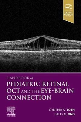
Cynthia A Toth
Handbook of Pediatric Retinal Oct and the Eye-Brain Connection
Cynthia A Toth
Handbook of Pediatric Retinal Oct and the Eye-Brain Connection
- Broschiertes Buch
- Merkliste
- Auf die Merkliste
- Bewerten Bewerten
- Teilen
- Produkt teilen
- Produkterinnerung
- Produkterinnerung
"Enhanced digital version included"--Cover.
Andere Kunden interessierten sich auch für
- Who Classification of Tumours of the Eye177,99 €
- Midwest MerchandiseTumbling Elephants: Animal Eye Chart11,99 €
- Steve VargoBut I Don't Sell: An Eye Care Professional's Guide to Being More Persuasive, Influential and Successful15,99 €
- Clinical Cases in Medical Retina133,99 €
- Jason HsuThe Pocket Guide to Medical Retina82,99 €
- Achilles Edward DavisHandbook of the Anatomy and Diseases of the Eye and Ear for Students and Practitioners26,99 €
- Joseph PanarelliThe Pocket Guide to Glaucoma83,99 €
-
-
-
"Enhanced digital version included"--Cover.
Hinweis: Dieser Artikel kann nur an eine deutsche Lieferadresse ausgeliefert werden.
Produktdetails
- Produktdetails
- Verlag: Elsevier Health Sciences
- Seitenzahl: 368
- Erscheinungstermin: 27. Juni 2019
- Englisch
- Abmessung: 155mm x 230mm x 10mm
- Gewicht: 570g
- ISBN-13: 9780323609845
- ISBN-10: 0323609848
- Artikelnr.: 57112572
- Verlag: Elsevier Health Sciences
- Seitenzahl: 368
- Erscheinungstermin: 27. Juni 2019
- Englisch
- Abmessung: 155mm x 230mm x 10mm
- Gewicht: 570g
- ISBN-13: 9780323609845
- ISBN-10: 0323609848
- Artikelnr.: 57112572
1 Introduction to OCT Imaging in Infants and Children
2 Basic Principles of OCT and OCTA Imaging of Infants and Children
3 Optimizing Systems and Setup for OCT and OCTA Imaging of Children and
Infants in the Nursery, Clinic, and Operating Room
4 OCT and OCTA Image Capture in the Nursery, Clinic, and Operating Room
5 Analyzing Structural Optical Coherence Tomography Images
6 Identifying Artifacts and Outliers in Structural Optical Coherence
7 Analyzing Optical Coherence Tomography Angiography
8 Identifying Artifacts in OCT Angiography
9 Introduction to Age-Dependent Features in Pediatric OCT Imaging
10 Foveal Development
11 Development of Retinal and Choroidal Vasculature and Peripheral
12 Vitreoretinal Abnormalities
13 Normal Optic Nerve Head: Anatomy and Development
14 Optic Nerve Head Abnormalities
15 Introduction to Research in Pediatric OCT Imaging
16 Considerations for Neonates and Children as a Vulnerable Research
Population
17 Benefit of Nondilated Imaging
18 Quality Assessment
19 Reproducibility of Imaging and Interpretation
20 Secure OCT Data Storage and Networking
21 Best Disease
22 Stargardt Disease (and Fundus Flavimaculatus)
23 Retinitis Pigmentosa
24 Other Forms of Retinitis Pigmentosa-Usher Syndrome, Leber Congenital
Amaurosis, and Bardet-Biedl Syndrome
25 Albinism
26 X-Linked Juvenile Retinoschisis
27 Other Inherited Retinal Diseases
28 Retinopathy of Prematurity
29 Familial Exudative Vitreoretinopathy and Norrie Disease
30 Incontinentia Pigmenti
31 Coats Disease and Coats Plus Syndrome
32 Sickle Cell Retinopathy
33 Epiretinal Membrane
34 Infectious Retinitis: TORCH Syndrome
35 White Dot Syndromes
36 Choroidal Granulomas: Tuberculosis and Sarcoidosis
37 Pediatric Intermediate Uveitis
38 Vogt-Koyanagi Harada Syndrome
39 Inflammatory and Idiopathic Choroidal Neovascularization
40 Nonaccidental Trauma
41 Ocular Injury
42 Traumatic Macular Hole
43 Commotio Retinae, Choroidal Rupture, and Sclopetaria
44 Retinal Laser Injury
45 Retinal Detachment and Proliferative Vitreoretinopathy
46 Retinoblastoma
47 Diffuse Choroidal Hemangioma
48 Circumscribed Choroidal Hemangioma
49 Choroidal Osteoma
50 Combined Hamartoma of the Retina and Retinal Pigment Epithelium
51 Retinal Astrocytic Hamartoma
52 Retinal Capillary Hemangioblastoma
53 Choroidal Nevus and Congenital Hypertrophy of the Retinal Pigment
Epithelium
54 Persistent Fetal Vasculature
55 Chorioretinal Coloboma
56 Myelinated Nerve Fiber Layer
57 Torpedo Maculopathy
58 Pathologic Myopia
59 Optic Nerve Pit
60 Optic Nerve Coloboma
61 Morning Glory
62 Optic Nerve Hypoplasia
63 Tilting of the Disc and Megalopapilla
64 Optic Atrophy
65 Optic Nerve Glioma
66 Papilledema and Disc Swelling Versus Traction Elevation
67 Optic Neuritis and Multiple Sclerosis
68 Neuroretinitis
69 Optic Nerve Head Drusen
70 Childhood Glaucoma