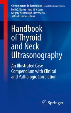
Incidentally discovered thyroid nodules and palpable neck masses present a common clinical problem. Further evaluation with neck ultrasound helps to characterize these lesions and stratify the risk of malignancy. Based on ultrasound characteristics, these lesions can be better defined using pathologic and other clinical data in order to determine the best management.
This practical handbook is a case compendium based on thyroid and neck ultrasonography patterns that are commonly encountered in clinical practice. A chapter will be designated for each of the major types of thyroid nodule ultrasound features, and additional chapters will be designated for other non-thyroid neck lesions. Beginning with imaging (photographic) examples of each of the ultrasound findings, clinical cases will be used to subdivide each feature and illustrate the differential diagnoses of the various types of thyroid and neck lesions. Associated cytology and histopathology images will beshown and described, as well as clinical data, including relevant history, physical exam, molecular marker results and clinical course. Chapters will include bulleted key points for quick reference as well as suggested readings for further investigation.
While several handbooks exist to educate readers about neck and thyroid ultrasonography and about cytopathology, this is a unique work that combines ultrasound and pathology data into a case-based, easy-to-read and concise format that fits in your pocket, adapting common terminology that is emerging for ultrasound, which will allow readers to employ a "standardized" approach to evaluating nodules, depending on the risk scoring system used.
Hinweis: Dieser Artikel kann nur an eine deutsche Lieferadresse ausgeliefert werden.
This book is an excellent resource with case-based chapters discussing all aspects of thyroid and parathyroid ultrasonography, thyroid fine needle aspiration and biopsy, thyroid cytopathology, and molecular marker testing. The concise chapters offer high-yield information authored by leaders in the field. This excellent handbook is a quick reference for those interested in thyroid and parathyroid disease. The chapters are well organized and easy to read, providing highlighted key points and suggested references for further exploration. The ultrasound images are exceptional, with numerous glossy photos highlighting the wide spectrum of thyroid and parathyroid pathology. The patient case correlation is especially helpful in maintaining clinical relevance for providers.
Shanika Samarasinghe, M.D.Loyola University Stritch School of Medicine
Shanika Samarasinghe, M.D.Loyola University Stritch School of Medicine

