- Broschiertes Buch
- Merkliste
- Auf die Merkliste
- Bewerten Bewerten
- Teilen
- Produkt teilen
- Produkterinnerung
- Produkterinnerung
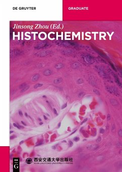
This book systematically illustrates theories and technologies in Histochemistry, including different kinds of enzymes, immunohistochemistry, polymerase chain reaction, related electron microscopic cytochemical techniques as well as the quantitative assay metrology. Abundant experiments as well as vivid images are demonstrated, making the book an essential reference for both graduate students and researchers in biochemistry.
Andere Kunden interessierten sich auch für
![Class 2 Transferases IV Class 2 Transferases IV]() A. Chang (Associate ed.)Class 2 Transferases IV149,99 €
A. Chang (Associate ed.)Class 2 Transferases IV149,99 €![Bioanalytics Bioanalytics]() Friedrich LottspeichBioanalytics75,99 €
Friedrich LottspeichBioanalytics75,99 €![Class 3.2 Hydrolases VIII Class 3.2 Hydrolases VIII]() Dietmar Schomburg / Ida Schomburg (eds.)Class 3.2 Hydrolases VIII229,99 €
Dietmar Schomburg / Ida Schomburg (eds.)Class 3.2 Hydrolases VIII229,99 €![Enzyme Handbook 13 Enzyme Handbook 13]() Enzyme Handbook 1339,99 €
Enzyme Handbook 1339,99 €![Class 3 Hydrolases Class 3 Hydrolases]() Dietmar Schomburg / Ida Schomburg (ed.) A. . Chang (Associate editor)Class 3 Hydrolases255,73 €
Dietmar Schomburg / Ida Schomburg (ed.) A. . Chang (Associate editor)Class 3 Hydrolases255,73 €![Lantibiotics and Related Peptides Lantibiotics and Related Peptides]() Ralph W. JackLantibiotics and Related Peptides115,99 €
Ralph W. JackLantibiotics and Related Peptides115,99 €![Class 2 Transferases Class 2 Transferases]() Dietmar Schomburg / Ida Schomburg (ed.) A. . Chang (Associate editor)Class 2 Transferases223,99 €
Dietmar Schomburg / Ida Schomburg (ed.) A. . Chang (Associate editor)Class 2 Transferases223,99 €-
-
-
This book systematically illustrates theories and technologies in Histochemistry, including different kinds of enzymes, immunohistochemistry, polymerase chain reaction, related electron microscopic cytochemical techniques as well as the quantitative assay metrology. Abundant experiments as well as vivid images are demonstrated, making the book an essential reference for both graduate students and researchers in biochemistry.
Hinweis: Dieser Artikel kann nur an eine deutsche Lieferadresse ausgeliefert werden.
Hinweis: Dieser Artikel kann nur an eine deutsche Lieferadresse ausgeliefert werden.
Produktdetails
- Produktdetails
- De Gruyter Textbook
- Verlag: De Gruyter
- 1. Auflage
- Seitenzahl: 244
- Erscheinungstermin: 21. August 2017
- Englisch
- Abmessung: 240mm x 170mm x 14mm
- Gewicht: 423g
- ISBN-13: 9783110524826
- ISBN-10: 3110524821
- Artikelnr.: 47554019
- Herstellerkennzeichnung
- de Gruyter Mouton
- Genthiner Straße 13
- 10785 Berlin
- productsafety@degruyterbrill.com
Jinsong Zhou, Xi¿an Jiaotong University, Shannxi Province, China
Contents
Chapter 1 Introduction
1.1 The contents and theories in Histochemistry
1.2 The basic requirements of Histochemistry methods
Chapter 2 The Tissue Preparation
2.1 Tissue collection
2.2 Fixation
2.3 Tissue washing-up, dehydration and clearance
2.4 Embedding
2.5 Sectioning
2.6 Adherence and mounting
2.7 Buffer
Chapter 3 The Carbohydrate and its Derivatives Histochemistry
3.1 Classification
3.2 Histochemistry methods
Chapter 4 The Nucleic Acid Histochemistry
4.1 DNA histochemical demonstration
4.2 Comparison demonstration of DNA and RNA Histochmistry
Chapter 5 The Lipid Histochemistry
5.1 Overview
5.2 Demonstration of lipids with physical methods
5.3 Demonstration of lipids with chemical methods
Chapter 6 Enzyme Histochemistry
6.1 Enzyme and its basic histochemical theory
6.2 Histochemistry for common enzymes
Chapter 7 The Basic Theory of Immunohistochemistry
7.1 Basic Immunology
7.2 Common markers and their detection
7.3 Basic conditions
Chapter 8 Common Methods Used in Immunohistochemistry
8.1 Principles
8.2 Immunofluorescence method
8.3 Immunoenzyme technique
8.4 Avidin-biotin method
8.5 Protein A method
8.6 Immunogold method and immune gold-silver method
Chapter 9 Specificity and Sensitivity of Immunohistochemistry
9.1 Specificity and specific staining
9.2 Control experiment
9.3 The methods to enhance immunohistochemical sensitivity
Chapter 10 The Double-Staining in Immunohistochemistry
10.1 Double-staining Immunohistochemistry on serial section
10.2 Immunofluorescence double labelling technique
10.3 Immunoenzyme double staining technique
10.4 Immunoenzyme-Immunofluorescence double staining
10.5 Immunoenzyme-Immunogold double staining
Chapter 11 The Lectin Histochemistry
11.1 Characteristics and application of lectin
11.2 Application of lectin Histochemistry
Chapter 12 The progress of in Situ Display
12.1 The envision method
12.2 The catalysed signal amplification method
12.3 The ferric oxide alternative method for HRP
Chapter 13 In Situ Hybridization Immunohistochemistry
13.1 Basic principle
13.2 Probe preparation
13.3 Procedure of in situ hybridization histochemistry
13.4 Factors affecting in situ hybridization
13.5 Control test
Chapter 14 In Situ Polymerase Chain Reaction Technique
14.1 Basic principle
14.2 Basic types
14.3 Procedure
14.4 Application of in situ PCR technology
Chapter 15 Electron Microscopic Cytochemical Technique
15.1 Electron microscopic enzyme cytochemistry technology
15.2 Electron microscopic Immunocytochemical technique
Chapter 16 The Quantitative Assay of Histochemistry Results
16.1 The photomicrography
16.2 The image analyzer
16.3 The flow cytometry
16.4 The laser scanning confocal microscopy
References

