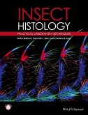
HISTOLOGICAL TECHNIQUE- A GUIDE FOR USE IN A LABORATORY COURSE IN HISTOLOGY by B. F. KINGSBURY. Originally published in 1927. PREFACE: The following represents the combination of technique notes written by the first author for use in connection with courses in Histology offered by him for medical, premedical, and veterinary students, with a similar outline of histological methods designed by the second author for use in courses dealing primarily with the histology of insects. Inasmuch as the methods-for the microscopic examination of animal structure are fundamentally the same, whether the struc ture is normal or pathological, the approach medical or zoological, it is believed that there has been here produced a book of much broader usefulness, without in any way sacrificing its value in histological work of more specific application. A rigid selection has been exercised, so that of the multitudinous methods employed in microscopic work only those are here given which meet the requirements for attaining a broad practical knowledge of animal structure. In special investigations it is necessary to make a study of the particular technical needs of the problem, and for this it is well to consult the larger works, of which may be mentioned: The Encyclopedia of Microscopic Technique [ 10]; The Microtomist's Vade-mecum, by A. B. Lee [ 35]; Mallory. and Wright, Pathological Technique [ 38]; Schu bcrg, A. [ 46]. These, as well as other books and articles, are listed in the Appendix, and reference is made to them in the text, either directly or by number [ in brackets]. Furthermore, in many in stances, direct reference to important papers is given in the text, thereby increasing the usefulness of the book for advanced stu dents in special fields. While the aim has been to present methods for the microscopic examination of any animal form, the emphasis is nevertheless placed on the technical needs of the premedical ( or medical) student and the student of Entomology. Contents include: INTRODUCTION vii FIXATION 1 Fixers, List of 6 ISOLATION 12 SECTIONING AND IMBEDDING 15 Schema for Imbedding 17 The Paraffin Method 18 The Ceiloidin Method 26 The Paraffin-Celloidin Method 33 The Freezing Method 35 STAINING 37 Stains, List of 41 Preparations for Staining 50 Schema for Staining 54 MOUNTING, SEALING AND LABELING 56 Slides and Covers 61 THE MICROSCOPE AND ACCESSORIES 63 SPECIAL METHODS 70 The Cell 70 Chitin 74 Connective Tissue 76 Muscle 81 The Nervous System 83 The Blood 95 Fine Injection 98 Silver Nitrate Impregnation 100 Histo-chemical Methods 101 SPECIAL METHODS FOR VARIOUS ANIMAL FORMS 107 Invertebrates in General 107 Arthropoda 118 Plankton Organisms 124 Taxonomic Material 126 APPENDIX 131 REFERENCES 133 INDEX 137. INTRODUCTION: Very few structures of the animal organism can be adequately examined microscopically without being first subjected to a pre paratory treatment involving in many cases the employment of complicated methods. Save in the case of the body fluids and certain membranes, animal tissues are bulky, more or less opaque, and therefore unsuited for examination under the microscope, which requires surface or thin layers of substance.
Bitte wählen Sie Ihr Anliegen aus.
Rechnungen
Retourenschein anfordern
Bestellstatus
Storno

