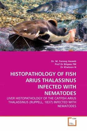
HISTOPATHOLOGY OF FISH ARIUS THALASSINUS INFECTED WITH NEMATODES
LIVER HISTOPATHOLOGY OF THE CATFISH ARIUS THALASSINUS (RUPPELL, 1837) INFECTED WITH NEMATODES
Versandkostenfrei!
Versandfertig in 6-10 Tagen
39,99 €
inkl. MwSt.

PAYBACK Punkte
20 °P sammeln!
The liver tissue damage in the catfish was observed. Lobular arrangement and central veins were obscured. Atrophy of liver tissue with large spaces in between was obvious. Portal tract areas were congested, hepatocyte cord arrangement and shape was disturbed. Section of nematode larvae apparently viable was present in liver tissue. Proliferation of the bile duct in portal tract area was obvious resulting into bile duct hyperplasia. Liver tissue appeared congested, loosing the usual arrangement of hepatocyte cord. Central veins were obliterated. Sinusoids were dilated due to atrophy of liver ce...
The liver tissue damage in the catfish was observed. Lobular arrangement and central veins were obscured. Atrophy of liver tissue with large spaces in between was obvious. Portal tract areas were congested, hepatocyte cord arrangement and shape was disturbed. Section of nematode larvae apparently viable was present in liver tissue. Proliferation of the bile duct in portal tract area was obvious resulting into bile duct hyperplasia. Liver tissue appeared congested, loosing the usual arrangement of hepatocyte cord. Central veins were obliterated. Sinusoids were dilated due to atrophy of liver cells. Mechanical damage of liver tissue due to nematode larval migration resulted into large spaces or tunnel around the larva. Liver cells arrangement was not identifiable. Hyperplasia of bile duct was a common finding in all the sections. Distinct egration of bile duct epithelium was also seen at some places. In the affected liver tissue ballooning of liver cells resulting into rounded outline instead of hexagonal shape was obvious. This information is useful for parasitologist, ichthyologist and fish farming agencies as well students in these field of studies.


