111,99 €
inkl. MwSt.
Versandkostenfrei*
Versandfertig in 6-10 Tagen
payback
56 °P sammeln
  • Broschiertes Buch

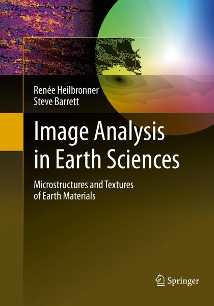
Image Analysis in Earth Sciences is a graduate level textbook for researchers and students interested in the quantitative microstructure and texture analysis of earth materials. Methods of analysis and applications are introduced using carefully worked examples. The input images are typically derived from earth materials, acquired at a wide range of scales, through digital photography, light and electron microscopy. The book focuses on image acquisition, pre- and post-processing, on the extraction of objects (segmentation), the analysis of volumes and grain size distributions, on shape fabric…mehr

Produktbeschreibung
Image Analysis in Earth Sciences is a graduate level textbook for researchers and students interested in the quantitative microstructure and texture analysis of earth materials. Methods of analysis and applications are introduced using carefully worked examples. The input images are typically derived from earth materials, acquired at a wide range of scales, through digital photography, light and electron microscopy. The book focuses on image acquisition, pre- and post-processing, on the extraction of objects (segmentation), the analysis of volumes and grain size distributions, on shape fabric analysis (particle and surface fabrics) and the analysis of the frequency domain (FFT and ACF). The last chapters are dedicated to the analysis of crystallographic fabrics and orientation imaging. Throughout the book the free software Image SXM is used.
Autorenporträt
Professor Renée Heilbronner has many years of experience in the field of image analysis and has developed several software packages for microstructure analysis for grain size, shape and strain determinations. She has also developed the CIP method for crystallographic texture determinations and orientation imaging based on polarization microscopy and digital image processing. She has contributed to the development of the freeware image analysis software (Image SXM, former NIH Image), and is a member of a growing group of international image analysis experts who are setting up workshops and building a network for microstructure and texture research involving mathematicians, material scientists and geologists. As an experienced teacher of image analysis at different levels ranging from general introductory courses to specialized texture workshops, she has taught at various universities all over the world.http://pages.unibas.ch/earth/micro Dr Steve Barrett is the author of the internationally renowned image analysis software Image SXM. He has been developing the software continuously over the past two decades, from its origins as a spin-off from the freeware NIH Image, to the extensions that allow it to handle images from over fifty types of optical and scanning microscopes. A customized version of this software (PrinCIPia) based on the CIP method can handle the calculation, display, analysis and manipulation of images representing the crystallographic orientation of grains in rock samples imaged by polarizing microscopes. He has published widely in the field of nanoscience and has also collaborated with medics to create microscopy image analysis software for medical applications (MIASMA). He has over twenty years experience teaching to undergraduates and postgraduates. http://www.liv.ac.uk/~sdb