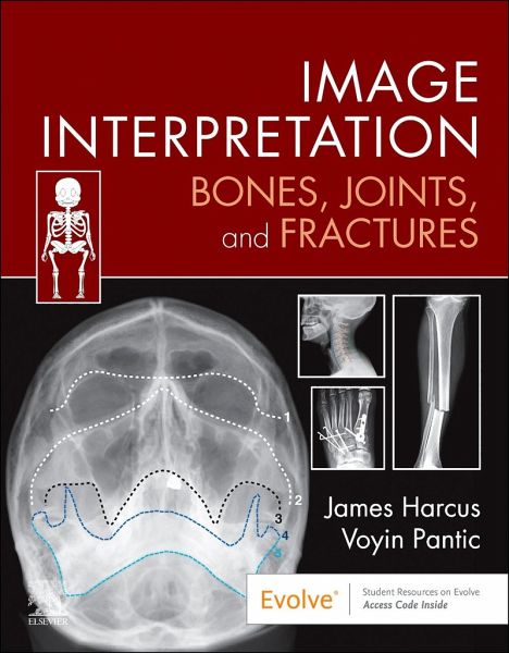
Interpreting X-ray images correctly is essential for diagnostic radiographers, as well as a widely used skill for emergency department doctors, nurse practitioners, and many other healthcare professions. This new title provides a systematic, methodical approach to musculoskeletal image interpretation and its role in the evaluation and treatment of injury. A companion to the eighth edition of Bones and Joints, this book covers the basic principles for interpreting images and then follows a simple regional approach to common radiographic projections. It goes on to consider common and important…mehr
Interpreting X-ray images correctly is essential for diagnostic radiographers, as well as a widely used skill for emergency department doctors, nurse practitioners, and many other healthcare professions. This new title provides a systematic, methodical approach to musculoskeletal image interpretation and its role in the evaluation and treatment of injury. A companion to the eighth edition of Bones and Joints, this book covers the basic principles for interpreting images and then follows a simple regional approach to common radiographic projections. It goes on to consider common and important fracture patterns and other injuries related to that region, as well as the differences between normal and abnormal images. Image Interpretation is an ideal learning guide for undergraduates, those transitioning to graduate roles or clinical practice, and other healthcare professionals wanting to supplement their training. User-friendly format: readers will be able to use the text to seamlessly explore the concepts between normal anatomy and abnormal radiographic appearances Systematic approach provided for each common radiographic projection Online case studies for readers to test and apply their clinical knowledge Key important learning points ('Insights') Annotated radiographic images and examples to support learningHinweis: Dieser Artikel kann nur an eine deutsche Lieferadresse ausgeliefert werden.
James Harcus qualified as a Diagnostic Radiographer from the University of Leeds in 2002 and worked at Leeds Teaching Hospitals NHS Trust as a radiographer across a range of specialities. In 2004 he began training in musculoskeletal image interpretation (reporting) at University of Bradford. After leaving Leeds in 2007 he took up a post in a large general practice surgery where he ran a small imaging service consisting of X-ray and ultrasound and during this time he completed his MSc in chest and abdominal image interpretation (reporting) and also his postgraduate certificate in musculoskeletal ultrasound back at the University of Leeds. After being a visiting and associate lecturer for a number of years, in 2012 he took up an academic post as a lecturer, and subsequently Senior Lecturer, at Sheffield Hallam University and became a fellow of the Higher Education Academy in 2014. In 2018 he helped establish the UK's first clinical academy for reporting radiographers. In December 2018 he moved to the University of Leeds as a lecturer in Diagnostic Imaging, teaching on both undergraduate and postgraduate programmes. He has a special focus on teaching skeletal anatomy which is ideal for this book.
Inhaltsangabe
¿1- Principles of Image Interpretation Theory of image viewing (e.g. forming and interpreting patterns); principles of image interpretation when viewing radiographic images (e.g. satisfaction of search, systematic approaches/patterns); types and formats of image interpretation (e.g. preliminary clinical evaluation/comment, clinical reporting); structuring a comment 2- Normal appearances of bones and joints Normal appearances of bones and joint; normal variants (including differentiating between normal variant and fracture). 3- Fractures and Joint Trauma Biomechanics of trauma; patterns of injury; fracture types and healing; treatment Chapters 4-9 will all follow a similar format by addressing each bone/joint in turn by considering: 1. * Systematic review of each bone/joint: how to assess, normal appearances/ measurements * Common normal variants * Patterns of injury * Common injuries; radiographic appearances supported by other modalities where appropriate 4- Upper limb Humerus, forearm, hand; elbow and wrist joints 5- Shoulder girdle and thorax Clavicle, scapula, sternum, and ribs; shoulder, acromioclavicular, and sternoclavicular joints 6- Lower limb Femur, tibia and fibula, foot; knee, ankle and foot joints 7- Pelvic girdle Hip bones; hip joint 8- Spine Cervical, thoracic, lumbar vertebrae; sacro-iliac joints 9- Facial bones and mandible 10- Practical applications of image interpretation CPD and audit of image interpretation practices. Role of artificial intelligence.
Es gelten unsere Allgemeinen Geschäftsbedingungen: www.buecher.de/agb
Impressum
www.buecher.de ist ein Internetauftritt der buecher.de internetstores GmbH
Geschäftsführung: Monica Sawhney | Roland Kölbl | Günter Hilger
Sitz der Gesellschaft: Batheyer Straße 115 - 117, 58099 Hagen
Postanschrift: Bürgermeister-Wegele-Str. 12, 86167 Augsburg
Amtsgericht Hagen HRB 13257
Steuernummer: 321/5800/1497
USt-IdNr: DE450055826