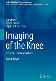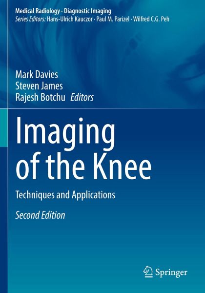
Imaging of the Knee
Techniques and Applications
Herausgegeben: Davies, Mark; James, Steven; Botchu, Rajesh
Versandkostenfrei!
Versandfertig in 6-10 Tagen
106,99 €
inkl. MwSt.

PAYBACK Punkte
53 °P sammeln!
This book is an entirely revised second edition of "Imaging of the Knee", published in 2003, and provides an up-to-date comprehensive review of imaging and pathologies of the knee. In the first part of the book, the various techniques employed when imaging the knee are discussed in detail. Individual chapters are devoted to radiography, arthrography, computed tomography and CT arthrography, magnetic resonance imaging and MR arthrography, and ultrasonography. The second part then documents the application of these techniques to the diverse clinical problems and diseases encountered in the knee....
This book is an entirely revised second edition of "Imaging of the Knee", published in 2003, and provides an up-to-date comprehensive review of imaging and pathologies of the knee.
In the first part of the book, the various techniques employed when imaging the knee are discussed in detail. Individual chapters are devoted to radiography, arthrography, computed tomography and CT arthrography, magnetic resonance imaging and MR arthrography, and ultrasonography. The second part then documents the application of these techniques to the diverse clinical problems and diseases encountered in the knee. Among the many topics addressed are congenital and developmental abnormalities, trauma, meniscal pathology, the cruciate and collateral ligaments, the postoperative knee, infection, arthritis, osteochondritis, osteonecrosis and tumors. Each chapter is written by an acknowledged expert in the field, and a wealth of illustrative material is included. This book will be of great value to radiologists and orthopaedic surgeons.
In the first part of the book, the various techniques employed when imaging the knee are discussed in detail. Individual chapters are devoted to radiography, arthrography, computed tomography and CT arthrography, magnetic resonance imaging and MR arthrography, and ultrasonography. The second part then documents the application of these techniques to the diverse clinical problems and diseases encountered in the knee. Among the many topics addressed are congenital and developmental abnormalities, trauma, meniscal pathology, the cruciate and collateral ligaments, the postoperative knee, infection, arthritis, osteochondritis, osteonecrosis and tumors. Each chapter is written by an acknowledged expert in the field, and a wealth of illustrative material is included. This book will be of great value to radiologists and orthopaedic surgeons.


