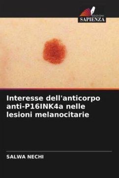
Interest of anti-P16INK4a antibody in melanocytic lesions
Versandkostenfrei!
Versandfertig in 6-10 Tagen
27,99 €
inkl. MwSt.

PAYBACK Punkte
14 °P sammeln!
The objective of our study is to evaluate the value of the expression of the p16INK4a antibody in the diagnosis of malignancy of melanocytic lesions. We compared the expression of this antibody between 25 melanomas and 25 common nevi, collected in the department of anatomy and pathological cytology of Mohamed Taher Maamouri Hospital, Nabeul during a period of 10 years (2010-2015). Nuclear staining was noted in 100% of nevi and in 13/25 (52%) of melanomas with a statistically significant difference (p
The objective of our study is to evaluate the value of the expression of the p16INK4a antibody in the diagnosis of malignancy of melanocytic lesions. We compared the expression of this antibody between 25 melanomas and 25 common nevi, collected in the department of anatomy and pathological cytology of Mohamed Taher Maamouri Hospital, Nabeul during a period of 10 years (2010-2015). Nuclear staining was noted in 100% of nevi and in 13/25 (52%) of melanomas with a statistically significant difference (p <0.0001). Cytoplasmic expression was not significantly different between nevi and melanomas. In nevi, an average of 54% of cells were positive with severe intensity (3+) versus an average of 12% in melanomas with low intensity. A threshold of positivity was defined by a percentage of labeled cells lower than 25% and a low intensity. Thus, the decrease or loss of P16 protein expression may constitute an argument to support the diagnosis of malignancy of melanocytic lesions. This argument must be confronted with the morphological data and other immunostaining.


