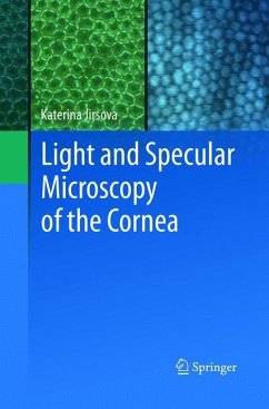
The atlas of the Light and Specular Microscopy of the Cornea, particularly of the corneal endothelium presents photographs of healthy and pathological corneas, as well as corneas prepared for grafting. Photographs are taken from donor or patient's corneas. The first part section of the atlas shows healthy corneas and its particular layers: the epithelium (superficial and basal cells, subepithelial nerve plexus), stroma and keratocytes, and the endothelium. Blood vessels or palisades of Vogt in limbus are shown as well. The second part section that shows corneas processed for grafting is focused focuses on the endothelial layer. Main causes of exclusion of corneas from grafting, such as the presence of dead cells, polymeghatism, pleomorphism, cornea guttata or stromal scars have been shown. The third part section of the atlas shows corneas before and after storage in tissue cultures or hypothermic conditions with the aim to assess its suitability of for tissue for grafting. Thelast final section contains photographs of pathological corneal explants
Bitte wählen Sie Ihr Anliegen aus.
Rechnungen
Retourenschein anfordern
Bestellstatus
Storno


