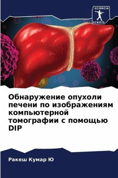
Liver Tumor Detection from CT Scan images using DIP
Versandkostenfrei!
Versandfertig in 6-10 Tagen
40,99 €
inkl. MwSt.

PAYBACK Punkte
20 °P sammeln!
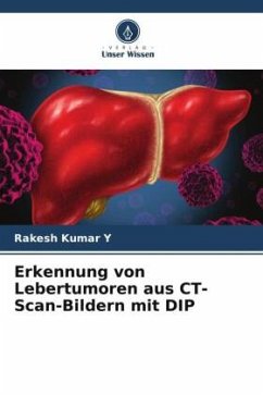
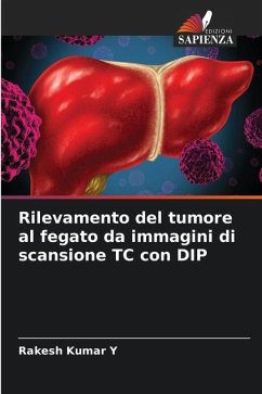
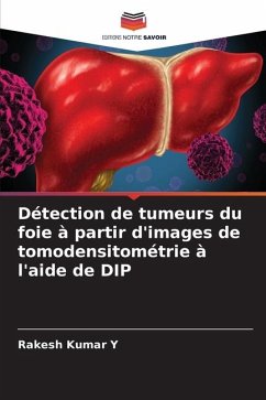
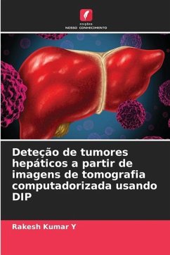
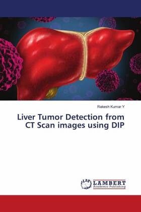
Liver imaging using abdominal CT images has been widely studied in the recent years and it is still a challenging task. Processing CT image includes the automatic diagnosis of liver and lesions part. Because of the high intensity similarity between liver tissues and nearby organs of liver it is difficult to segment liver and tumor. Segmentation of extracted region as an imaging biomarker forms an essential component of "Radiomics". This book presents brief intro to liver tumor in CT Scan images, different methods in detecting liver tumor, automatic liver tumor segmentation from abdominal CT sc...
Liver imaging using abdominal CT images has been widely studied in the recent years and it is still a challenging task. Processing CT image includes the automatic diagnosis of liver and lesions part. Because of the high intensity similarity between liver tissues and nearby organs of liver it is difficult to segment liver and tumor. Segmentation of extracted region as an imaging biomarker forms an essential component of "Radiomics". This book presents brief intro to liver tumor in CT Scan images, different methods in detecting liver tumor, automatic liver tumor segmentation from abdominal CT scan images is presented. A statistical parameter-based approach is used to distinguish liver tumor tissue from other abdominal organs. The existing segmentation methods such as region growing and intensity based thresholding methods are investigated and compared with statistical parameter-based method.


