Nicht lieferbar

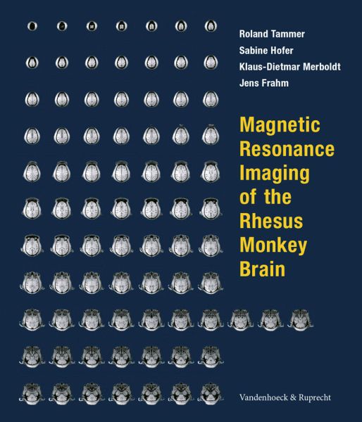
Magnetic Resonance Imaging of the Rhesus Monkey Brain
. Rhesus Monkey Brain
Mitwirkender: Tammer, Roland; Merboldt, Klaus-Dietmar; Hofer, Sabine
Versandkostenfrei!
Nicht lieferbar
Magnetic resonance imaging (MRI) of the central nervous system plays an increasingly important role in the neurosciences involving humans and animals. Nonhuman primates are of special relevance because of their immunological, physiological, and behavioral similarities to humans. Therefore, the lack of detailed anatomical MRI data of the rhesus monkey brain was a strong motivation for this work. It provides the first comprehensive in vivo MRI atlas of the living macaque brain at the highest technical quality currently available. Three-dimensional coverage of the brain in horizontal, coronal, an...
Magnetic resonance imaging (MRI) of the central nervous system plays an increasingly important role in the neurosciences involving humans and animals. Nonhuman primates are of special relevance because of their immunological, physiological, and behavioral similarities to humans. Therefore, the lack of detailed anatomical MRI data of the rhesus monkey brain was a strong motivation for this work. It provides the first comprehensive in vivo MRI atlas of the living macaque brain at the highest technical quality currently available. Three-dimensional coverage of the brain in horizontal, coronal, and sagittal sections at unprecedented 0.5 mm isotropic spatial resolution is at the core of the atlas with an indication of anatomical structures. Multiple contrasts are supplied compatible with human MRI. Advanced techniques such as magnetic resonance angiography and diffusion tensor imaging are exploited for a visualization of the intracranial vasculature and the virtual reconstruction of nerve fiber tracts, respectively. "Readability" for the non-expert is ensured by a simple introduction into the principles and applications of MRI. Detailed descriptions cover all aspects of animal handling, experimental procedures, and image presentation in a stereotaxic coordinate system. The atlas is expected to serve as a reference source for easy identification of anatomical structures in the rhesus monkey brain. It is a "must" on the desktop of primatologists and neuroscientists in a broad range of disciplines.


