Andrew R. Houghton (United Lincolnshire Ho Consultant Cardiologist
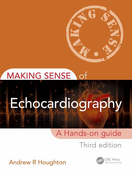
Making Sense of Echocardiography
A Hands-on Guide
Andrew R. Houghton (United Lincolnshire Ho Consultant Cardiologist
Making Sense of Echocardiography
A Hands-on Guide
- Broschiertes Buch
- Merkliste
- Auf die Merkliste
- Bewerten Bewerten
- Teilen
- Produkt teilen
- Produkterinnerung
- Produkterinnerung
Building on the success of the second edition, the third edition of Making Sense of Echocardiography: A Hands-on Guide provides a timely overview for those learning echocardiography for the first time as well as an accessible handbook that experienced sonographers can refer to.
Andere Kunden interessierten sich auch für
- Cecil BosherLandmarks In Cardiac Surgery267,99 €
- Imaging in Critical Care Medicine81,99 €
- Richard A Jonas (Children's Na Children's National Heart InstituteComprehensive Surgical Management of Congenital Heart Disease430,99 €
- David MaisuradzeEchocardiography in Metabolic Syndrome68,99 €
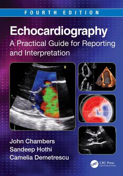
- Camelia Demetrescu (Guyâ s and St Thomasâ Hospitals, London, UK)Echocardiography64,99 €
- Problem-Based Obstetric Ultrasound61,99 €
- Kshitij MankadRevision Notes for the Final FRCR Part A68,99 €
-
-
-
Building on the success of the second edition, the third edition of Making Sense of Echocardiography: A Hands-on Guide provides a timely overview for those learning echocardiography for the first time as well as an accessible handbook that experienced sonographers can refer to.
Hinweis: Dieser Artikel kann nur an eine deutsche Lieferadresse ausgeliefert werden.
Produktdetails
- Produktdetails
- Making Sense of
- Verlag: Taylor & Francis Ltd
- 3 ed
- Seitenzahl: 306
- Erscheinungstermin: 21. August 2023
- Englisch
- Abmessung: 177mm x 255mm x 19mm
- Gewicht: 752g
- ISBN-13: 9781032303543
- ISBN-10: 1032303549
- Artikelnr.: 67826403
- Making Sense of
- Verlag: Taylor & Francis Ltd
- 3 ed
- Seitenzahl: 306
- Erscheinungstermin: 21. August 2023
- Englisch
- Abmessung: 177mm x 255mm x 19mm
- Gewicht: 752g
- ISBN-13: 9781032303543
- ISBN-10: 1032303549
- Artikelnr.: 67826403
Andrew Houghton MA(Oxon), DM, BM BCh, FRCP (Lond) Consultant Cardiologist and Clinical Head of Echocardiography, Grantham and District Hospital, Grantham, Lincolnshire, UK Dr Andrew R. Houghton studied medicine at the University of Oxford and undertook postgraduate training in Nottingham and Leicester. He was appointed as consultant cardiologist at Grantham & District Hospital in Lincolnshire, UK, in 2002. His subspecialty interest is in non-invasive cardiac imaging, in particular echocardiography and cardiovascular MRI. He has been a member of the British Society of Echocardiography's departmental accreditation committee, and a lecturer at BSE annual congresses. Dr Houghton has co-authored a number of textbooks, including Making Sense of the ECG (now in its fifth edition) and its companion volume Making Sense of the ECG: Cases for Self-Assessment, and is also a faculty member at the Medmastery online medical education website. Making Sense of the ECG has won several awards, including the Royal Society of Medicine's Richard Asher prize and the British Medical Association's Student Textbook Award, while Making Sense of Echocardiography was highly commended at the BMA Medical Book Awards.
Preface
Acknowledgements
Author biography
Acronyms and abbreviations
Part 1 Essential principles
1. History of echocardiography
2. Cardiac anatomy and physiology
3. Physics and instrumentation
4. Doppler physics
5. Myocardial mechanics
6. Service provision
Part 2 Cardiac imaging techniques
7. The standard transthoracic echo study
8. Transoesophageal echo
9. Stress echo
10. Contrast echo
11. Tissue Doppler imaging
12. Speckle tracking
13. 3D echo
14. Intravascular ultrasound and epicardial echo
15. Alternative cardiac imaging techniques
Part 3 Clinical cases
16. The left ventricle and its systolic function
17. Coronary artery disease and regional left ventricular function
18. Left ventricular diastolic function
19. The left atrium
20. The aortic valve
21. The mitral valve
22. The right ventricle
23. The right atrium
24. The tricuspid valve
25. The pulmonary valve
26. Pulmonary hypertension
27. Heart valve repair and replacement
28. Endocarditis
29. The cardiomyopathies
30. The pericardium
31. The aorta
32. Cardiac masses
33. Congenital heart disease
34. Common echo requests
Appendix - Echo resources
Index