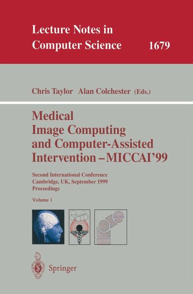
Medical Image Computing and Computer-Assisted Intervention - MICCAI'99
Second International Conference, Cambridge, UK, September 19-22, 1999, Proceedings
Herausgegeben: Taylor, Chris; Colchester, Alan

PAYBACK Punkte
20 °P sammeln!
This book constitutes the refereed proceedings of the Second International Conference on Medical Image Computing and Computer-Assisted Intervention, MICCAI'99, held in Cambridge, UK, in September 1999.The 133 revised full papers presented were carefully reviewed and selected from a total of 213 full-length papers submitted. The book is divided into topical sections on data-driven segmentation, segmentation using structural models, image processing and feature detection, surfaces and shape, measurement and interpretation, spatiotemporal and diffusion tensor analysis, registration and fusion, visualization, image-guided intervention, robotic systems, and biomechanics and simulation.
Abnormal white matter of the brain is common to patients with one of s- eral di?erent diseases (including multiple sclerosis (MS) and Alzheimer s d- ease (AD)) and also appears in normal (asymptomatic) aging (NA) subjects. Better characterization of the nature of these white matter changes can help to improve our understanding of the biological processes at work. Clinically, it is interesting to be able to di?erentiate between di?erent disease states and to ?nd markerswhich allowearlydiagnosis.Conventionalspin echo magnetic resonance imaging is sensitive to these white matter changes. MRI studies of patients and volunteershaveindicatedthatthepatternsofbrainchangeassociatedwiththese processes are di?erent. An important goal is to be able to quantitatively study these di?erences. Many automated and semi-automated segmentation algorithms for quan- tatively assessing these brain changes have been developed and validated. Most of these algorithms have aimed at determining a binary characterization of each voxelas one of a groupof possible tissue classes.This approachhas been limited bytwofactors.First,abnormalwhitematter isoftenisointensewithnormalgrey matter and previous studies have been limited by the inability to discriminate between some abnormal white matter and normal grey matter [1,2]. Secondly, white matter damage appears as an heterogenous region of abnormal signal - tensity but binarization of the segmentation treats all levels of signal intensity abnormality equally.


