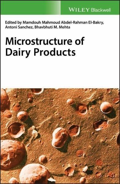
Microstructure of Dairy Products
Herausgeber: El-Bakry, Mamdouh; Mehta, Bhavbhuti M; Sanchez, Antonio
Microstructure of Dairy Products
Herausgeber: El-Bakry, Mamdouh; Mehta, Bhavbhuti M; Sanchez, Antonio
- Gebundenes Buch
- Merkliste
- Auf die Merkliste
- Bewerten Bewerten
- Teilen
- Produkt teilen
- Produkterinnerung
- Produkterinnerung
Provides the most recent developments in microscopy techniques and types of analysis used to study the microstructure of dairy products This comprehensive and timely text focuses on the microstructure analyses of dairy products as well as on detailed microstructural aspects of them. Featuring contributions from a global team of experts, it offers great insight into the understanding of different phenomena that relate to the functional and biochemical changes during processing and subsequent storage. Structured into two parts, Microstructure of Dairy Products begins with an overview of…mehr
Andere Kunden interessierten sich auch für
- A. A. AwadTechnology of Cereal-rich Probiotic Dairy Beverage38,99 €
- Handbook of Drying for Dairy Products233,99 €
- Dairy Science and Technology Handbook, Volume 1356,99 €
- Dairy Science and Technology Handbook, Volume 2356,99 €
- Dairy Science and Technology Handbook, Volume 3356,99 €
- Ahmed EmamMozzarella and its analogues: manufacture and functionality63,99 €
- Dairy Powders and Concentrated Products319,99 €
-
-
-
Provides the most recent developments in microscopy techniques and types of analysis used to study the microstructure of dairy products This comprehensive and timely text focuses on the microstructure analyses of dairy products as well as on detailed microstructural aspects of them. Featuring contributions from a global team of experts, it offers great insight into the understanding of different phenomena that relate to the functional and biochemical changes during processing and subsequent storage. Structured into two parts, Microstructure of Dairy Products begins with an overview of microscopy techniques and software used for microstructural analyses. It discusses, in detail, different types of the following techniques, such as: light microscopy (including bright field, polarized, and confocal scanning laser microscopy) and electron microscopy (mainly scanning and transmission electron microscopy). The description of these techniques also includes the staining procedures and sample preparation methods developed. Emerging microscopy techniques are also covered, reflecting the latest advances in this field. Part 2 of the book focuses on the microstructure of various dairy foods, dividing each into sections related to the microstructure of milk, cheeses, yogurts, powders, and fat products, ice cream and frozen dairy desserts, dairy powders and selected traditional Indian dairy products. In addition, there is a review of the localization of microorganism within the microstructure of various dairy products. The last chapter discusses the challenges and future trends of the microstructure of dairy products. * Presents complete coverage of the latest developments in dairy product microscopy techniques * Details the use of microscopy techniques in structural analysis * An essential purchase for companies, researchers, and other professionals in the dairy sector Microstructure of Dairy Products is an excellent resource for food scientists, technologists, and chemists--and physicists, rheologists, and microscopists--who deal in dairy products.
Hinweis: Dieser Artikel kann nur an eine deutsche Lieferadresse ausgeliefert werden.
Produktdetails
- Produktdetails
- Verlag: Wiley
- Seitenzahl: 400
- Erscheinungstermin: 22. Oktober 2018
- Englisch
- Abmessung: 246mm x 175mm x 25mm
- Gewicht: 957g
- ISBN-13: 9781118964224
- ISBN-10: 1118964225
- Artikelnr.: 51802865
- Verlag: Wiley
- Seitenzahl: 400
- Erscheinungstermin: 22. Oktober 2018
- Englisch
- Abmessung: 246mm x 175mm x 25mm
- Gewicht: 957g
- ISBN-13: 9781118964224
- ISBN-10: 1118964225
- Artikelnr.: 51802865
Mamdouh Mahmoud Abdel-Rahman El-Bakry is a Postdoctoral Researcher in the Department of Chemical Engineering at the Universitat Autònoma of Barcelona, Spain. Antoni Sanchez is a Professor in the Department of Chemical Engineering at the Universitat Autònoma of Barcelona, Spain. Bhavbhuti M. Mehta is an Associate Professor in the Dairy Chemistry Department, SMC College of Dairy Science, Anand Agricultural University, Gujarat, India.
List of Contributors xiii Preface xv 1 Microscopy Techniques for Dairy
Products - An Introduction 1 Mark A.E. Auty 1.1 Introduction 1 1.1.1 Brief
History and Background 1 1.2 Conventional Optical Microscopy Techniques 4
1.2.1 Conventional Light Microscopy - Optical Contrast 4 1.2.1.1 Bright
Field 4 1.2.1.2 Polarized Light 4 1.2.1.3 Phase Contrast 4 1.2.1.4
Differential Interference Contrast 5 1.2.1.5 Fluorescence 5 1.2.2 Chemical
Contrast Techniques in Light Microscopy 5 1.3 Confocal Scanning Laser
Microscopy 6 1.3.1 Confocal Principle 6 1.3.2 Identifying Dairy Primary
Components in CSLM: Labeling Strategies 8 1.3.2.1 Generic Labeling 8
1.3.2.2 Specific Labeling 10 1.3.2.3 Covalent Labeling 11 1.3.3 Some
Applications of Confocal Microscopy to Dairy Products and Ingredients 12
1.3.3.1 Spreads 12 1.3.3.2 Emulsions and Foams 12 1.3.3.3 Fermented Milks
12 1.3.3.4 Cheese 13 1.3.3.5 Dairy Powders 13 1.3.3.6 Milk Protein Gel
Systems 14 1.3.3.7 Dynamic CSLM Techniques 14 1.4 Electron Microscopy (EM)
Techniques 16 1.4.1 Transmission Electron Microscopy 16 1.4.2 Scanning
Electron Microscopy 18 1.4.3 Other EM Techniques 18 1.4.3.1 X-ray
Microanalysis 18 1.4.3.2 Cryo-electron Microscopy 19 1.4.3.3 Environmental
and Variable Pressure SEM 20 1.5 Emerging Microscopy Techniques 20 1.5.1
Atomic Force Microscopy 20 1.5.2 Advanced Fluorescence Microscopy
Techniques 22 1.5.3 Confocal Raman Microscopy 22 1.5.4 X-ray
Nano/Microtomography 22 1.5.5 Super-Resolution Microscopy 23 1.6 Image
Analysis 23 1.7 Conclusions 24 References 24 2 Light Microscopy and CSLM
Techniques, Principles and Applications 33 Johan Hazekamp 2.1 Introduction
33 2.1.1 The History of Microscopy 33 2.1.2 Evolution of Confocal
Microscopy 34 2.1.3 Food Microscopy 35 2.1.4 Wide Field Microscopy 36 2.1.5
Confocal Scanning Laser Microscopy (CSLM) 38 2.2 Sample Preparation and
Specific Staining and Labeling 41 2.2.1 Specific Labeling 44 2.2.2 Dynamic
Imaging 46 2.2.3 Future Perspectives 46 References 47 3 Electron Microscopy
Techniques 51 Semih Otles and Vasfiye Hazal Ozyurt 3.1 Introduction 51 3.2
Types of EM 51 3.2.1 Scanning Electron Microscopy (SEM) 51 3.2.2
Transmission Electron Microscopy (TEM) 52 3.2.3 Cryo-SEM 52 3.2.4 Cryo-TEM
53 3.2.5 Environmental Scanning Electron Microscopy (ESEM) 53 3.3 Sample
Preparation for EMs 53 3.3.1 Scanning Electron Microscopy 53 3.3.2
Transmission Electron Microscopy 53 3.3.3 Cryo-Scanning Electron Microscopy
53 3.4 Dairy Microstructure 54 3.5 Electron Microscopy for the Dairy
Product 54 3.6 Summary 60 References 64 4 Emerging Techniques for
Microstructural Analysis 67 I. Hernando, E. Llorca, and A. Quiles 4.1
Introduction 67 4.2 Scanning Probe Microscopy 67 4.2.1 Scanning Tunneling
Microscope (STM) 69 4.2.2 Atomic Force Microscope (AFM) 70 4.2.3
Applications of the Main Probe Microscopes 71 4.3 X-Ray Tomography 72 4.4
Small-Angle-Scattering (SAS) Methods: SAXS and SANS 74 4.4.1 Small-Angle
X-Ray Scattering (SAXS) 74 4.4.2 Small-Angle Neutron Scattering (SANS) 75
4.4.3 Applications of Small-Angle-Scattering Methods 75 4.5 Vibrational
Spectroscopies (Fourier Transform Infrared-FTIR and Raman Microscopy) 75
4.5.1 Fourier Transform Infrared (FTIR) Spectroscopy 76 4.5.2 Raman
Spectroscopy 78 4.6 Magnetic Resonance: NMR and MRI 80 4.7 Conclusions 82
References 82 5 Quantitative Image Analysis in Microscopy 89 Gaetano Impoco
5.1 Aim and Scope 89 5.2 Image Analysis Software 90 5.3 Applications to
Microscopy for Dairy Science 97 5.3.1 Porosity 98 5.3.2 Fat Globules 99
5.3.3 Microbial Cells 100 5.4 Image Analysis and Quantitative Measurement
100 5.4.1 Image Analysis Basics 101 5.4.1.1 Feature Detection 102 5.4.1.2
Quantitative Analysis 103 5.4.2 Common Pitfalls 105 5.4.3 Misuse and Wrong
Interpretation of Image Analysis Results 114 5.4.4 Good Practices 116 5.4.5
Image Analysis in your Lab 119 5.5 Conclusions 122 Acknowledgments 123
References 123 6 Microstructure of Milk 127 Michael H. Tunick 6.1
Components of Milk 127 6.2 Fat 127 6.2.1 Fat Globules 127 6.2.2 Milkfat
Globule Membrane 128 6.2.3 Cream 129 6.3 Protein 133 6.3.1 Types of Protein
133 6.3.2 Casein Micelles in Bovine Milk 133 6.3.3 Casein Micelles in
Caprine Milk 133 6.3.4 Casein Micelles in Milk of Other Species 136 6.3.5
Micelle Structure 136 6.4 Bacteria and Somatic Cells 137 6.5 Concentrated
Milk 138 6.6 Digested Milk 140 6.7 Conclusion 142 Acknowledgments 142
References 142 7 Microstructure of Cheese Products 145 Bhavbhuti M. Mehta
7.1 Introduction 145 7.2 Factors Affecting the Development of
Microstructures in Cheeses 146 7.2.1 Addition of Calcium Chloride 148 7.2.2
Rennet Coagulation 149 7.2.3 Acid-Coagulation 150 7.2.4 Coagulation
Temperature 150 7.2.5 Syneresis 151 7.2.6 Salting 151 7.2.7 Ripening 152
7.2.8 Homogenization and High Pressure Treatments 153 7.2.9 Evaporation and
Ultrafiltration Treatments 155 7.2.10 Freezing 156 7.2.11 Fat Replacers 156
7.3 Microstructures of Various Components in Cheese Matrix 158 7.3.1
Protein in Cheese Matrix 158 7.3.2 Fat Globule in Cheese Matrix 159 7.3.3
Calcium in Cheese Matrix 162 7.4 Crystals in Cheese Matrix 162 7.5 Starter
Bacteria in Cheese Matrix 163 7.6 Microstructure of Selected Varieties of
Cheeses 164 7.6.1 Processed Cheese 164 7.6.1.1 Curd Granules and Fat 166
7.6.1.2 Occurrence of Crystals 166 7.6.2 Cheese Analogs 166 7.6.3 Feta
Cheese 167 7.6.4 Domiati Cheese 167 7.6.5 Fresh Cheese 167 7.6.6 Cream
Cheese 168 7.6.7 Mold-Ripened Cheeses 169 7.6.8 Cheese Powder 169 7.7
Cheese Matrix and Digestion 170 7.8 Conclusions 171 References 171 8
Microstructural Aspects of Yogurt and Fermented Milk 181 P.H.P. Prasanna,
C.S. Ranadheera, and J.K. Vidanarachchi 8.1 Yogurt and Fermented Milk: An
Overview 181 8.2 Yogurt and Fermented Milk: Production Technologies 184 8.3
Microstructure of Yogurt and Fermented Milk 187 8.4 Factors Influencing
Microstructure of Yogurt and Fermented Milk 188 8.4.1 Effects of Type of
Milk on Structure 188 8.4.2 Rate of Inoculation Level and Starter Culture
Composition on Microstructure of Yogurt and Fermented Milk 189 8.4.2.1 Rate
of Inoculation 189 8.4.2.2 Culture Composition 189 8.4.3 Effect of
Exopolysaccharide Producing Starter Culture on Microstructure 190 8.4.4
Incubation Temperature on Structure 191 8.4.5 Effect of Different
Processing Steps 191 8.4.5.1 Homogenization of Milk 191 8.4.5.2 Heat
Treatment of Milk 192 8.4.5.3 Effect of Stirring 192 8.4.6 Effect of
Addition of Different Hydrocolloids and Fibers on Microstructure 193 8.5
Microscopy Methods Used for Analyzing Microstructure of Fermented Milk 194
8.5.1 Light Microscopy 194 8.5.1.1 Bright Field Light Microscopy 194
8.5.1.2 Polarized Light Microscopy 194 8.5.1.3 Fluorescence Microscopy 195
8.5.1.4 Confocal Laser Scanning Microscopy 197 8.5.2 Electron Microscopy
198 8.5.2.1 Scanning Electron Microscopy 198 8.5.2.2 Transmission Electron
Microscopy 200 8.6 Conclusions 201 References 202 9 Microstructure of Milk
Fat and its Products 209 Pere Randy R. Ramel and Alejandro G. Marangoni 9.1
Introduction 209 9.2 Milk Fat Crystal Structure 211 9.2.1 Mesoscale
Structure of Milk Fat 211 9.2.1.1 Polymorphism 211 9.2.1.2 Phase Behavior
and Fractionation 213 9.2.1.3 Solid Fat Content and Crystallization/Melting
Behavior 214 9.2.2 Nanoscale Structure of Fat Crystal Networks 216 9.3
Effect of Different Factors on the Crystallization Behavior and
Microstructure of Milk Fat 218 9.3.1 Processing Conditions 218 9.3.1.1
Different Crystallization Mechanisms 218 9.3.1.2 Crystallization
Temperature and Cooling Rate 218 9.3.1.3 Agitation, Shear and Ultrasound
219 9.3.2 Composition 220 9.3.2.1 Minor Components 220 9.3.2.2 Blending
with Different Fats and Oils, and Waxes 220 9.3.3 In a Dispersed State
(Emulsion) 221 9.3.3.1 Emulsified State (Cream) vs Bulk State or Anhydrous
Milk Fat (AMF) 222 9.3.3.2 Emulsion Droplet Size 222 9.3.3.3 Addition of
Emulsifiers 223 9.3.4 In Food Matrices 223 9.3.4.1 Water-in-Oil Emulsion
223 9.3.4.2 Foamed Emulsions 224 9.3.4.3 Chocolate 225 9.3.4.4 Cheese 226
9.4 Impact of Resulting Microstructure on the Properties of Different Milk
Fat Products 226 9.4.1 Rheology 226 9.4.2 Thermal Stability 229 9.4.3
Sensory Qualities 229 9.5 Conclusions 229 References 230 10 Microstructure
of Ice Cream and Frozen Dairy Desserts 237 Samantha R. VanWees and Richard
W. Hartel 10.1 Overview of Frozen Desserts 237 10.1.1 Ingredients 238
10.1.2 Processing 239 10.2 Frozen Dessert Structure 240 10.2.1 Serum Phase
240 10.2.2 Ice Crystals 242 10.2.3 Fat Phase 245 10.2.4 Air Cells 247
10.2.5 Proteins and Hydrocolloids 250 10.3 Storage 251 10.3.1
Recrystallization 251 10.3.2 Sugar Crystallization 253 10.3.3 Air
Coarsening 254 10.3.4 Shrinkage 255 10.4 Conclusion 256 References 256 11
Whey Wastes and Powders 261 J. Chandrapala 11.1 Whey 261 11.2 Current Whey
Uses 263 11.3 Processing of Liquid Whey 263 11.3.1 Recovery of Casein Fines
and the Separation of Fat 264 11.3.2 Concentration of Total Solids 265
11.3.3 Drying 266 11.3.4 Fractionation of Total Solids 270 11.4 Whey
Powders 274 11.4.1 Whey Protein Concentrates 275 11.4.2 Whey Protein
Isolates 277 11.4.3 Whey Protein Hydroxylates 279 11.4.4 Other Whey Powders
280 11.4.4.1 Defatted Whey Protein Concentrates 280 11.4.4.2 Demineralized
Whey Protein Concentrates 280 11.4.4.3 Delactosed Whey Powders 283 11.4.4.4
Acid Whey Powders 283 11.4.4.5 Salty Whey Powders 284 11.5 Utilization and
Applications of Whey Powders 285 11.6 Conclusion 287 References 287 12
Microstructure of Selected Traditional Indian Dairy Products 293 Bhavbhuti
M. Mehta 12.1 Introduction 293 12.2 Heat Desiccated Dairy Products 294
12.2.1 Khoa and Khoa-Based Sweets 294 12.2.1.1 Microstructure of Khoa 294
12.2.1.2 Microstructure of Gulabjamun 295 12.2.1.3 Microstructure of Burfi
and Kalakand 298 12.3 Heat-Desiccated Milk Cereal Based Desserts 299 12.3.1
Microstructure of Kheer 299 12.4 Heat-Acid Coagulated Dairy Products 300
12.4.1 Microstructure of Paneer 300 12.4.1.1 Fried Paneer 300 12.4.2
Microstructure of Chhana and Chhana Based Sweets 302 12.4.2.1
Microstructure of Rasogolla 302 12.4.2.2 Microstructure of Chhana Podo 305
12.5 Fermented Dairy Products 306 12.5.1 Microstructure of Dahi 306 12.5.2
Microstructure of Shrikhand 306 12.6 Conclusion 307 References 307 13 Using
Microscopy for Microorganism Localization within Dairy Products 311 I.T.
Smykov 13.1 Introduction 311 PART 1 312 13.1.1 Microorganisms and Starters
312 13.1.2 Techniques Used in the Microstructure Analyses 313 13.1.3
Interactions Occurring in the Microstructure 315 PART 2 318 13.2 Materials
and Methods 318 13.2.1 Bacterial Strains and Dairy Products 318 13.2.2
Electron Microscopy 318 13.2.2.1 Surface Topography Heavy Metal Shadowing
319 13.2.2.2 Negative Staining Transmission Electron Microscopy 319 13.2.3
Freeze-Fracture Replication 319 13.3 Results and Discussion 320 13.3.1
Casein Micelle 320 13.3.2 Bacteria 324 13.3.3 Bacteria in a Protein Matrix
327 13.3.4 Bacteria in Cheese Eyes 331 13.3.5 Bacteria in Yoghurt 333
13.3.6 Bacteriophages 336 13.4 Conclusions 338 Acknowledgment 339
References 339 14 Microstructure of Dairy Products: Challenges and Future
Trends 345 Maricê Nogueira de Oliveira 14.1 Introducing Microstructure of
Dairy Products 345 14.2 Microstructure of Fermented Milks 346 14.3
Microstructure of Yogurt and Milk Drinks 347 14.3.1 Yogurt 347 14.3.2 Milk
Drinks or Lactic Beverages 354 14.4 Microstructure of Cheeses 356 14.5
Conclusion 359 References 359 Index 363