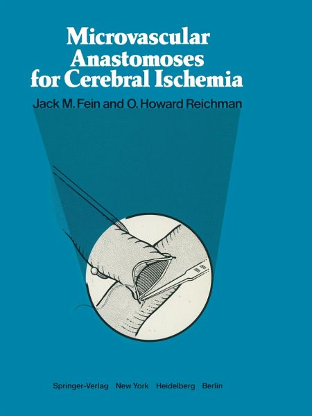
We have witnessed a remarkable development during the past 10 years in the development of extra-intracranial anastomosis to revascularize the brain. Initially, the intention was to create ameans of performing embolectomy in small corticaI arteries after cardiac surgery. Gradually a plan was conceived to form an extra-intracranial bypass(10) to treat inaccessible lesions of the carotid and vertebral arteries as weIl as tumors and giant aneurysms that involved these arteries. The basic techniques and principles of microvascular sur gery, which had been refined over 30 years on peripheral ar…mehr
We have witnessed a remarkable development during the past 10 years in the development of extra-intracranial anastomosis to revascularize the brain. Initially, the intention was to create ameans of performing embolectomy in small corticaI arteries after cardiac surgery. Gradually a plan was conceived to form an extra-intracranial bypass(10) to treat inaccessible lesions of the carotid and vertebral arteries as weIl as tumors and giant aneurysms that involved these arteries. The basic techniques and principles of microvascular sur gery, which had been refined over 30 years on peripheral ar teries(1-7, 9) were applied in 1966 to the intracerebral arteries of dogs(4). The arterial patehing and suturing were successful, while extra-intracraniallong bypasses were a constant failure. This was attributed to the damage that was inevitably inflicted on the vasa vasorum of the autogenous graft during dissec tion(9). This situation posed a dilemma until eventually the idea of Pool and Potts(8) was adopted and an anastomosis was performed on a dog between the superficiaI temporaI artery and a cortical branch. Because of the vesseIs' small size and the doubts regarding its capacity for improving the circulation, the procedure was performed with a certain amount of apprehen sion. It was a pleasant surprise, therefore, to discover that it was possible to attain a high rate of patency. If the middle cerebral artery was us ed and was isolated from its carotid in flow, an even higher rate was achieved.Hinweis: Dieser Artikel kann nur an eine deutsche Lieferadresse ausgeliefert werden.
I. Structure and Function.- 1. Anatomy and Physiology Pertinent to Stroke.- 2. Canine Cerebral Ischemia.- 3. Anatomical Studies of the Posterior Circulation Relevant to Occipital Artery Bypass.- 4. Comparison of Blood Flow and Patency in Arterial and Vein Grafts to the Basilar Artery.- 5. The Endothelial Surface of Arteries: A Scanning Electron Microscopic Examination of Normal and Anastomosed Vessels.- 6. The Experimental Effects of Prostaglandin E1 on Small Vessel Patency.- II. Metabolism.- 7. Oxidative Metabolism in Cerebral Ischemia.- 8. Evidence for Barbiturate Protection in Focal Cerebral Ischemia: A Hypothesis for Mechanism and Clinical Utility.- 9. Transient Ischemic Attacks and Metabolic Aspects of Their Relief by Microneurosurgical Anastomosis.- III. Clinical Evaluation and Diagnosis.- 10. Management of Risk Factors and Other Diseases in Candidates for Microneurosurgical Anastomosis in Cerebral Ischemia.- 11. Ocular Plethysmography and Suction Ophthalmo-dynamometry in the Diagnosis of Carotid Occlusive Disease.- 12. Collateral Circulation of the Brain.- 13. Angiographic Cerebral Blood Flow Patterns in STA-MCA Anastomosis Candidates.- IV. Surgical Techniques.- 14. Contemporary Techniques of Cerebral Revascularization.- 15. External Carotid-Middle Cerebral Artery Bypass Using Free Graft Bypass.- 16. Electromagnetic Blood Flowmetry in Microvascular Anastomosis.- 17. Brain Vascularization by Transplanted Omentum.- V. Clinical-Hemodynamic Considerations.- 18. Augmentation of Collateral Hemispheric Blood Pressure following Superficial Temporal to Middle Cerebral Artery Anastomosis: Documentation by Ocular Plethysmography.- 19. Regional Cerebral Blood Flow Studies following Superficial Temporal-Middle Cerebral Artery Anastomosis.- 20. Current Status of Regional Cerebral Blood Flow Measurement in Revascularization Microsurgery of the Brain.- 21. Estimation of Flow Through STA Bypass Graft.- 22. Cerebral Blood Flow in Stroke-Type Patients.- VI. Clinical Results.- 23. Progression of Middle Cerebral Artery Stenosis to Occlusion Without Symptoms following Superficial Temporary Artery Bypass: Case Report.- 24. Experiences with the STA-Cortical MCA Anastomosis in 46 Cases.- 25. Clinical Experiences with STA-MCA Anastomosis in 54 Cases.- 26. Microneurosurgical Arterial Anastomoses in Patients with Prolonged Reversible Ischemic Neurological Deficits (PRIND).- 27. Review of Experience with 50 Consecutive Cases of Superficial Temporal Artery to Middle Cerebral Artery Anastomosis for Treatment of Cerebrovascular Occlusive Disease.- VII. Other Applications.- 28. Microvascular Anastomosis and Carotid Artery Ligation for Fibromuscular Hyperplasia and Carotid Artery Aneurysm.
Es gelten unsere Allgemeinen Geschäftsbedingungen: www.buecher.de/agb
Impressum
www.buecher.de ist ein Internetauftritt der buecher.de internetstores GmbH
Geschäftsführung: Monica Sawhney | Roland Kölbl | Günter Hilger
Sitz der Gesellschaft: Batheyer Straße 115 - 117, 58099 Hagen
Postanschrift: Bürgermeister-Wegele-Str. 12, 86167 Augsburg
Amtsgericht Hagen HRB 13257
Steuernummer: 321/5800/1497
USt-IdNr: DE450055826